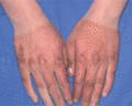
Figure 101.1
Erythropoietic protoporphyria showing marked purpura with sharp cut‐off after sunlight exposure.
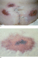
Figure 101.2
(a) Heparin necrosis at sites of subcutaneous heparin injection. (b) Close‐up of a 15 cm lesion on the left abdomen. The lesion is a non‐palpable haem...
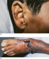
Figure 101.3
Cold‐induced lesions due to cryofibrinogenaemia, (a) on the ear and (b) on the foot. An acral location is typical for cryogelling. The foot lesion sho...
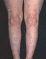
Figure 101.4
Sneddon syndrome showing a typical, broad, racemose livedo patterning.
