Figure 102.1
Cutaneous small‐vessel vasculitis producing palpable purpura. (Courtesy of Andrew Carmichael.)
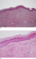
Figure 102.5
Leukocytoclastic (small vessel) vasculitis at (a) lower and (b) higher magnifications. There is visible vascular wall damage with evidence of red bloo...
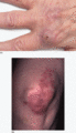
Figure 102.9
Erythema elevatum diutinum. (a) On the hands. (b) Early non‐fibrotic lesions at a typical site on the knee. This patient also had EED on the hands, an...
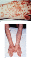
Figure 102.13
IgA vasculitis. (a) Haemorrhagic vesicles present on the hand. (Courtesy of Andrew Carmichael, South Tees Hospitals NHS Trust, UK.) (b) Vasculitis ex...
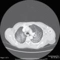
Figure 102.17
Computed tomography of the thorax demonstrating pulmonary haemorrhage affecting both lung fields in microscopic polyangiitis.
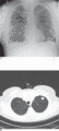
Figure 102.21
Granulomatosis with polyangiitis. (a) X‐ray showing bilateral nodules.(b) Computed tomography of the thorax demonstrating a thick‐walled cavity in the...
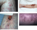
Figure 102.25
Cutaneous polyarteritis nodosa. (a) Erythematous lesions on the leg. (b) Nodules and ulceration on the leg. (c) Ulcerating lesions on the leg. (d) Liv...
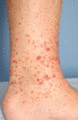
Figure 102.2
Cutaneous small‐vessel vasculitis demonstrating a haemorrhagic vesicle. (Courtesy of Andrew Carmichael.)
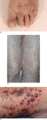
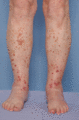
Figure 102.6
Cutaneous small‐vessel vasculitis (CSVV). (a) Vesicles in a dependent area on the foot. (b) Purpura on the thighs; there was similar involvement on th...
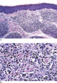
Figure 102.10
Granuloma faciale. (a) Low‐magnification view showing perivascular nodular infiltrates within the dermis. (b) At a higher magnification the infiltrate...
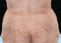
Figure 102.14
Cryoglobulinaemic vasculitis.
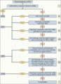
Figure 102.18
Classification of antineutrophil cytoplasmic antibody vasculitides and polyarteritis nodosa (PAN). ACR, American College of Rheumatology; CHCC, Chapel...
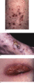
Figure 102.22
Granulomatosis with polyangiitis. (a) Ulcerated lesions of cutaneous small‐vessel vasculitis. (b) Larger ulcerated lesions with background vasculitis....
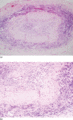
Figure 102.26
Giant cell arteritis at (a) lower and (b) higher magnifications. There are obliterative vascular changes with a lymphocytic and multinucleated giant c...
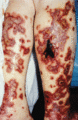
Figure 102.3
Ulcerated necrotic lesions in a livedo distribution suggestive of medium‐vessel disease. (Courtesy of Andrew Carmichael.)
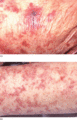
Figure 102.7
Vasculitis due to sepsis, in a patient with impaired level of consciousness. The necrotic lesion in (a) and the reticulate pattern on the leg in (b) a...
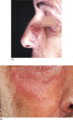
Figure 102.11
Granuloma faciale. (a) Reddish brown plaque on the nose (courtesy of Dr G. Dawn, Monklands Hospital, UK). (b) Close up of a facial plaque.
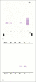
Figure 102.15
Serum protein electrophoresis. (a) Demonstrating a clear band in lanes 1, 2 and 5. Lanes 3 and 4 are negative controls and lane 5 is a urine control f...
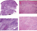
Figure 102.19
Granulomatosis with polyangiitis. (a) There is extensive leukocytoclastic vasculitis involving the entire dermis. (b) Note the extensive area of colla...
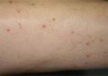
Figure 102.23
Relatively subtle vasculitis on the legs in eosinophilic granulomatosis with polyangiitis. The patient also had eosinophilia and rapidly developed a m...
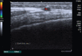
Figure 102.27
Colour Doppler ultrasound of the temporal artery demonstrating vessel wall oedema (the halo sign).
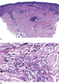
Figure 102.4
Leukocytoclastic vasculitis. (a) Low‐magnification photomicrograph showing perivascular infiltrates and fibrinoid deposits within the vessels of the u...
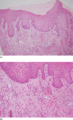
Figure 102.8
Erythema elevatum diutinum at (a) lower and (b) higher magnifications. There is a perivascular infiltrate containing neutrophils, with leukocytoclasis...
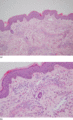
Figure 102.12
IgA vasculitis at (a) lower and (b) higher magnifications. There is perivascular leukocytoclasis and fibrin deposition, and eosinophils are present. ...
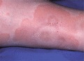
Figure 102.16
Urticarial vasculitis.
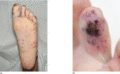
Figure 102.20
Granulomatosis with polyangiitis. (a) Cutaneous infarction. (b) Digital infarction.
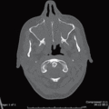
Figure 102.24
Computed tomography scan of paranasal sinuses demonstrating opacification of the maxillary sinuses and bone erosion in eosinophilic granulomatosis wit...
