Figure 108.1
Anatomical landmarks of the auricle.

Figure 108.5
Microtia – absent ear canal, partly formed pinna and additional cartilage remnant and skin tag. (Source: Dr Michael Saunders, Consultant Paediatric Ot...

Figure 108.9
Keloids following ear piercing.

Figure 108.13
Angiolymphoid hyperplasia with eosinophilia. Firm red‐brown nodules at the entrance to the external auditory canal.

Figure 108.17
Juvenile spring eruption. Subtle oedematous plaques on helix of ear.

Figure 108.21
Otitis externa. (a) The pinna and skin nearby is erythematous with crusting and scaling. The entrance to the canal is narrowed. (b) The pinna is eryth...

Figure 108.25
Atypical fibroxanthoma. A firm fleshy tumour of the pinna.

Figure 108.2
Coarse terminal hair on the auricle: a trait possibly associated with the Y chromosome.

Figure 108.6
Treacher Collins syndrome – marked retrognathia, low set pinna with microtia. (Source: Dr Michael Saunders, Consultant Paediatric Otolaryngologist, Br...

Figure 108.10
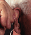
Chondrodermatitis nodularis of the helix. A superficially ulcerated, exquisitely tender nodule.

Figure 108.14
Subepidermal calcifying nodule. Hard whitish nodule with slight overlying scale.

Figure 108.18
Porphyria cutanea tarda. Firm whitish sclerodermoid changes at the site of repeated blistering.

Figure 108.22
Discoid lupus erythematosus . Erythema with adherent scaling in the concha.

Figure 108.26
(a) B‐cell lymphoma presenting as a purple nodular swelling in the retroauricular fold. (b) Erythematous swelling of the ear lobe due to infiltration ...

Figure 108.3
Coloboma of the eye, heart defects, atresia of the nasal choanae, retardation of growth and/or development, genital and/or urinary abnormalities, and ...

Figure 108.7
Pre‐auricular sinus.

Figure 108.11
Pseudocyst. Asymptomatic fluctuant swellings on the upper pinna.

Figure 108.15
Gouty tophi. Yellowish dermal nodules.

Figure 108.19
Psoriasis. (a) Well‐defined erythema with prominent scaling at entrance to ear canal – close‐up in (b).

Figure 108.23
Chronic otitis externa. There is longstanding eczematous change, in part due to contact allergic reactions to ear drops, and induration causing narro...

Figure 108.27
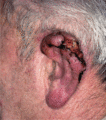
Squamous carcinoma of the auricle. An advanced tumour with extensive destruction of the ear cartilage. (Courtesy of Mr D. Baldwin, Southmead Hospital...

Figure 108.4
(a) Branchio‐oto‐renal syndrome: second arch branchial cyst (arrowed), pre‐auricular skin tag, pre‐auricular sinus and microtia with absent ear canal....

Figure 108.8
Diagonal earlobe crease in an infant with Beckwith–Wiedemann syndrome.

Figure 108.12
Alkaptonuria. The auricular cartilage has a distinctive blue colour. (Courtesy of Dr P. Hollingworth, Southmead Hospital, Bristol, UK.)

Figure 108.16
Cutaneous lupus erythematosus. Acute erythema and erosions following sun exposure.

Figure 108.20
Several firm white ‘weathering’ nodules on the helical rim.

Figure 108.24
Basal cell carcinoma of the external auditory canal. An erythematous tumour presenting as obstruction at the entrance of the canal. (Courtesy of Mr M...

Figure 108.28
Squamous carcinoma of the external auditory canal. Purulent discharge, inflammation and destruction of the meatus. (Courtesy of Mr D. Baldwin, Southm...

