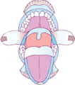
Figure 110.1
Diagram of the oral cavity.
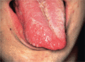
Figure 110.5
Lymphangioma of the tongue: a common site.
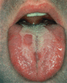
Figure 110.9
Classical geographic tongue (lingual erythema migrans).
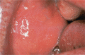
Figure 110.13
Fordyce spots: sebaceous glands in the buccal mucosa.
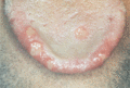
Figure 110.17
Major aphthous ulcers.
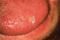
Figure 110.21
Aphthous‐like ulceration in HIV disease.
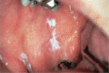
Figure 110.25
Lichen planus: plaque‐like lesions resemble leukoplakia.
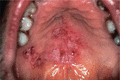
Figure 110.29
Chronic oral lesions in discoid lupus erythematosus.
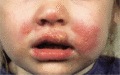
Figure 110.33
Primary herpetic stomatitis with extraoral lesions.
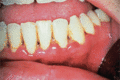
Figure 110.37
Acute necrotizing gingivitis showing typical ulceration of interdental gingival papillae. This was in HIV infection.
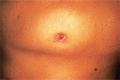
Figure 110.41
Sinus on the chin related to a dental abscess on a mandibular incisor tooth.
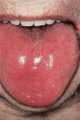
Figure 110.45
Atrophic glossitis in vitamin B 12 deficiency.
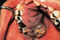
Figure 110.49
Kaposi sarcoma in a typical site with a characteristic purplish appearance. (Courtesy of Dr J.B. Epstein, Cancer Control Agency, Vancouver, Canada.)
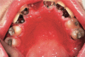
Figure 110.53
Denture‐induced stomatitis showing diffuse erythema in the denture‐bearing area.
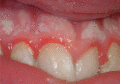
Figure 110.57
Materia alba.
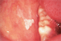
Figure 110.61
Homogeneous leukoplakia in the buccal mucosa.
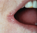
Figure 110.65
Angular cheilitis.
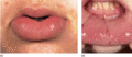
Figure 110.69
Granulomatous cheilitis of the lower lip. (Courtesy of Addenbrooke's Hospital, Cambridge, UK.)
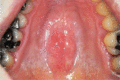
Figure 110.2
Torus palatinus.
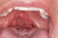
Figure 110.6
Lingual tonsil showing a well‐demarcated midline groove. (Courtesy of Dr C.T.C. Kennedy, Bristol Royal Infirmary, Bristol, UK.)
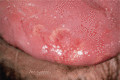
Figure 110.10
Somewhat less obvious signs of lingual erythema migrans.
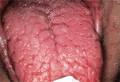
Figure 110.14
Fissured or scrotal tongue.
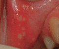
Figure 110.18
Herpetiform ulceration
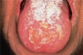
Figure 110.22
Herpes simplex lingual recurrence, and candidosis in leukaemia: similar lesions may be seen in HIV infection.
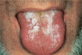
Figure 110.26
Lichen planus on the tongue.
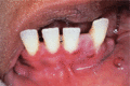
Figure 110.30
Pemphigoid: vesicles and desquamative gingivitis.
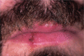
Figure 110.34
Herpes labialis.
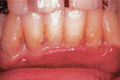
Figure 110.38
Untreated acute necrotizing gingivitis can lead to extensive gingival ulceration and irreparable damage.
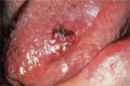
Figure 110.42
Macroglossia and oral petechiae in amyloidosis.
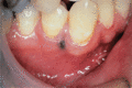
Figure 110.46
Amalgam tattoo in a common site. This was presumably related to filling of the deciduous predecessor.
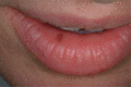
Figure 110.50
Melanotic macule of the lower lip.
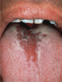
Figure 110.54
Median rhomboid glossitis.
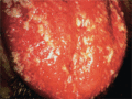
Figure 110.58
Thrush: scattered white lesions on an erythematous background.
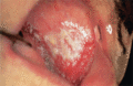
Figure 110.62
Speckled leukoplakia.
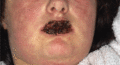
Figure 110.66
Haemorrhagic crusting of the lips in Stevens–Johnson syndrome.
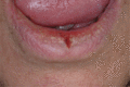
Figure 110.70
Lip fissure.
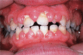
Figure 110.3
Gingival hyperplasia in phenytoin therapy. Concomitant folate deficiency in this patient also caused mouth ulcers, seen in the maxillary buccal vestib...
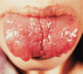
Figure 110.7
Multiple neuromas of the lips and tongue in a patient with multiple endocrine neoplasia syndrome (type 2). (Courtesy of Dr M. Hartog, Bristol Royal I...
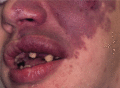
Figure 110.11
Haemangioma affecting the lip in Sturge–Weber syndrome.

Figure 110.15
Angular sinus (lip‐pit), a congenital anomaly.
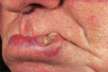
Figure 110.19
Squamous cell carcinoma of the lip.
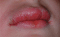
Figure 110.23
Orofacial granulomatosis.
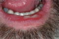
Figure 110.27
Erosive lichen planus.
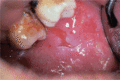
Figure 110.31
Pemphigus vulgaris: irregular persistent oral erosions.
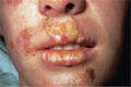
Figure 110.35
Impetigo.
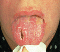
Figure 110.39
Gumma.
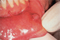
Figure 110.43
Mucocoele.
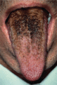
Figure 110.47
Black hairy tongue.
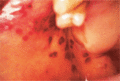
Figure 110.51
Oral purpura in thrombocytopenia.
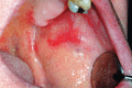
Figure 110.55
Erythroplasia.
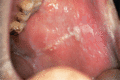
Figure 110.59
Frictional keratosis and cheek biting (morsicatio buccarum) at the occlusal line.
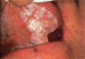
Figure 110.63
Sublingual keratosis.
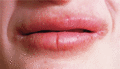
Figure 110.67
Factitious cheilitis due to repeated lip sucking.
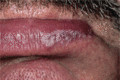
Figure 110.71
Discoid lupus erythematosus of the lower lip.
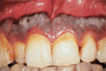
Figure 110.4
Gingival hyperpigmentation of racial origin. The white lesion is due to accumulated oral debris – oral hygiene is very poor.
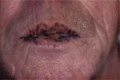
Figure 110.8
Peutz–Jeghers syndrome.
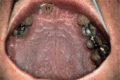
Figure 110.12
Darier disease: oral white lesions resemble those of nicotinic stomatitis.
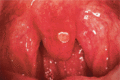
Figure 110.16
Recurrent aphthae.
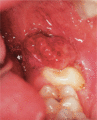
Figure 110.20
Oral squamous cell carcinoma.
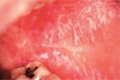
Figure 110.24
Lichen planus: reticulopapular lesions in the common oral site, the buccal mucosa.
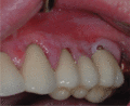
Figure 110.28
Lichen planus on the gingivae
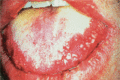
Figure 110.32
Scattered ulcers and a furred tongue in primary herpetic stomatitis.
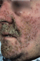
Figure 110.36
Eczema herpeticum.
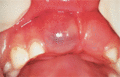
Figure 110.40
Bluish, fluctuant swelling of an oral cyst, in this case an eruption cyst over an erupting maxillary permanent incisor. (The lesion on the maxillary c...
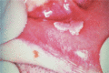
Figure 110.44
Warts on the lower lip in HIV infection. (There is also a healing herpes simplex lesion on the lip.)
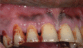
Figure 110.48
Betel staining of teeth.
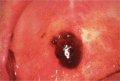
Figure 110.52
Angina bullosa haemorrhagica: a large blood blister in a typical site on the soft palate. The adjacent whitish lesions are from scarring after a previ...
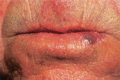
Figure 110.56
Venous lake of the lip. (Courtesy of Addenbrooke's Hospital, Cambridge, UK.)
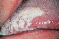
Figure 110.60
Hairy leukoplakia. Found mainly in HIV infection, vertical white ridges on the lateral margin of the tongue.
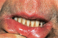
Figure 110.64
Chronic actinic cheilitis with leukoplakia. (Courtesy of Addenbrooke's Hospital, Cambridge, UK.)
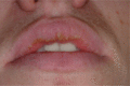
Figure 110.68
Exfoliative cheilitis.
