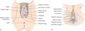
Figure 112.1
(a) The vulva. (b) The clitoris. (From Neill and Lewis 2009 [ ]. Reproduced with permission of John Wiley & Sons.)
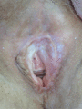
Figure 112.5
Lichen sclerosus showing white sclerotic plaques and architectural change.
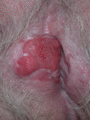
Figure 112.9
Squamous cell carcinoma arising on a background of lichen sclerosus.
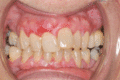
Figure 112.13
Gingival erythema in vulvo‐vaginal–gingival syndrome.
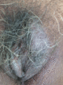
Figure 112.17
Lichen simplex.
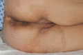
Figure 112.21
Tinea cruris.
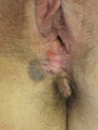
Figure 112.25
Vulval intraepithelial neoplasia.
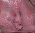
Figure 112.29
Tear of the right inferior aspect of the nymphohymenal sulcus.
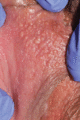
Figure 112.2
Fordyce spots.
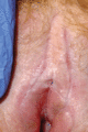
Figure 112.6
Fissuring in lichen sclerosus.
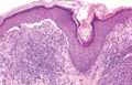
Figure 112.10
Histological features of lichen planus.
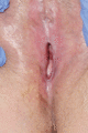
Figure 112.14
Scarring in erosive vulval lichen planus.
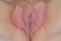
Figure 112.18
Vulval psoriasis.
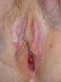
Figure 112.22
Vulval warts with plaques in the interlabial sulci.
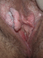
Figure 112.26
Verrucous carcinoma on a background of lichen sclerosus.
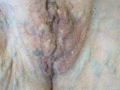
Figure 112.3
Vulval varicosities.
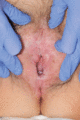
Figure 112.7
Thickened epithelium in acanthotic lichen sclerosus.
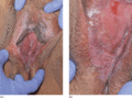
Figure 112.11
(a) Classic vulval lichen planus showing plaques in the interlabial sulci. (b) Classic vulval lichen planus with Wickham striae.
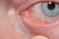
Figure 112.15
Lacrimal duct scarring in erosive lichen planus.
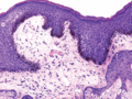
Figure 112.19
Histological features of vulval melanosis.
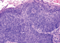
Figure 112.23
Histological features of undifferentiated vulval intraepithelial neoplasia.
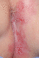
Figure 112.27
Extramammary Paget disease
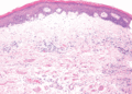
Figure 112.4
Histological features of lichen sclerosus.
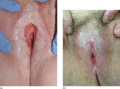
Figure 112.8
(a) Scarring in lichen sclerosus with ecchymosis. (b) Scarring in lichen sclerosus with loss of the labia minora and sealing of the clitoral hood.
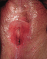
Figure 112.12
Lichen planus: vulval aspect showing glazed erythema and distortion of the architecture, with a remnant of the left labium minus and buried clitoris a...
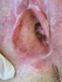
Figure 112.16
Chronic vulval purpura.
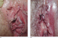
Figure 112.20
(a) In situ melanoma. (b) Vulval melanosis.
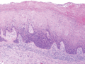
Figure 112.24
Histological features of differentiated vulval intraepithelial neoplasia.
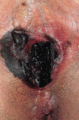
Figure 112.28
Melanoma of the vulva. (Courtesy of Dr F. A. Ive, Durham, UK.)
