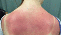
Figure 127.1
Erythema following acute UV radiation (intense natural sunlight) exposure (‘sunburn’).
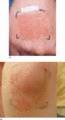
Figure 127.5
Artificially provoked polymorphic light eruption (PLE). (a) Back of the hand after iterative UVA photoprovocation (2 × 25 J/cm 2 ). (b) Erythema multi...
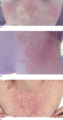
Figure 127.9
Papules and plaques on photo‐exposed sites during an acute flare of actinic prurigo (AP). (a) On the cheeks and nose; note distal nose involvement. (b...

Figure 127.13
Histopathology of chronic actinic dermatitis. (a) Epidermal spongiosis, acanthosis and an upper dermal lymphohistiocytic inflammatory infiltrate (seen...
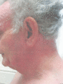
Figure 127.17
This patient presented with a photo‐exposed site dermatitis but phototesting was normal and this was photoaggravated seborrhoeic dermatitis (Courtesy...
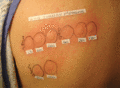
Figure 127.21
Abnormal immediate photosensitivity to UVA (365 nm) and visible (400 and 430 nm) wavelengths in a patient with treatment‐resistant solar urticaria. No...
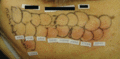
Figure 127.25
Phototoxicity to demeclocycline. There is predominant UVA and visible light photosensitivity. Acute erythema, oedema and urticaria were seen at the te...
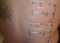
Figure 127.29
Positive photopatch testing to sunscreen chemicals.
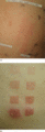
Figure 127.33
Abnormal erythemal responses on (a) narrow‐band UVB (TL‐01) MED, and (b) psoralen–UVA (PUVA) MPD testing.
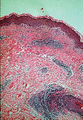
Figure 127.2
Histopathology of polymorphic light eruption showing a superficial and deep, predominantly perivascular, lymphohistiocytic, dermal inflammatory infilt...
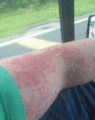
Figure 127.6
Polymorphic light eruption provoked on the extensor forearm by window glass‐transmitted light (patient's own photograph).
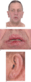
Figure 127.10
Persistent actinic prurigo in an adult. (a) Note inflammation of the lower eyelids, distal nose involvement and cheilitis (with coincidental xanthelas...
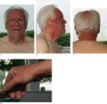
Figure 127.14
Chronic actinic dermatitis presenting acutely with a photo‐exposed‐site dermatitis. (a, b) Note the prominent head and neck involvement and sharp cut‐...
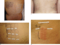
Figure 127.18
Abnormal monochromator phototesting in chronic actinic dermatitis (CAD) showing broad‐band UVB, UVA and visible light photosensitivity. (a) This patie...
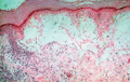
Figure 127.22
Histopathology of hydroa vacciniforme showing epidermal necrosis, dermal oedema and a lymphohistiocytic inflammatory infiltrate.
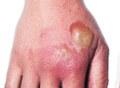
Figure 127.26
Phytophotodermatitis. Erythema and blistering is seen on sites immersed in psoralen‐rich lime marinade during barbeque preparations and subsequently e...
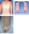
Figure 127.30
(a) Photoaggravated guttate psoriasis. Note the sparing at photoprotected sites under the crop top. (b) This patient had atopic eczema and systemic lu...
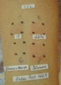
Figure 127.34
Abnormal erythemal responses on phototesting to compact fluorescent lamps. Note the negative response at the site irradiated through Dermagard film, w...
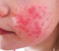
Figure 127.3
Papulovesicular polymorphic light eruption in a child; facial involvement is seen more commonly in children.
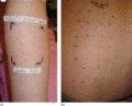
Figure 127.7
Artificially provoked papular polymorphic light eruption. (a) On the extensor forearm (two UVA exposures: 10 and 20 J/cm 2 ). (b) At narrow‐band UVB (...
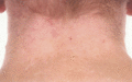
Figure 127.11
Scarring and hypopigmentation on the chronically photo‐exposed site of the back of the neck in actinic prurigo.
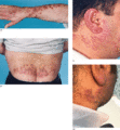
Figure 127.15
In patients of higher skin phototypes (IV to VI) with chronic actinic dermatitis, a nodular prurigo‐like morphology may be apparent. (a) Prominent inv...
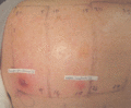
Figure 127.19
Strong positive patch tests in a patient with chronic actinic dermatitis. Patch testing is an essential investigation to undertake in this patient gro...
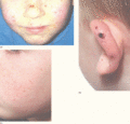
Figure 127.23
Hydroa vacciniforme. (a) Papules, vesiculation and haemorrhagic crusting on the cheeks (patient's own photograph). (b) Papules on the ears. (c) Subtle...
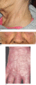
Figure 127.27
Chlorpromazine phototoxicity. (a) Characteristic brown‐grey pigmentation associated with chronic photosensitivity secondary to chlorpromazine. (b) Vio...
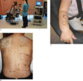
Figure 127.31
(a) Monochromator phototesting using a fibreoptic light guide. (b, c) Phototesting and other photo‐investigations should be possible in most children....
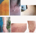
Figure 127.4
Polymorphic light eruption (PLE) subtypes. (a) Mixed papulovesicular PLE in an adult with vesicles predominating. (b) Plaque PLE showing variably size...
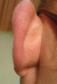
Figure 127.8
Juvenile spring eruption: sunlight‐induced papules on the prominent pinna of a young male.
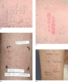
Figure 127.12
Abnormal phototesting in actinic prurigo (AP). (a) Monochromator phototesting showing broad‐band UVB and UVA photosensitivity. (b) A positive UVA phot...
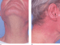
Figure 127.16
This patient presented with unexplained erythroderma. Slight sparing is seen (a) under the chin and (b) at the collar line and within the scalp. Chron...
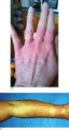
Figure 127.20
Solar urticaria. (a) Acute erythema on the back of the hand after 2 min of wintertime daylight exposure. Note sparing of the distal phalanges and unde...
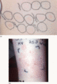
Figure 127.24
(a) Monochromator phototesting showing abnormal UVA photosensitivity. (b) Positive UVA photoprovocation in hydroa vacciniforme.
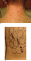
Figure 127.28
Voriconazole photosensitivity. (a) Abnormal stellate lentigines developed on photo‐exposed sites in this patient who had been taking voriconazole for ...
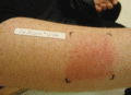
Figure 127.32
Iterative photoprovocation, usually using a broad‐band UVA source, can be particularly useful in confirming a diagnosis of polymorphic light eruption,...
