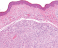
Figure 137.1
Fibrous papule with hyalinized collagen bundles and increased dilated vascular channels.

Figure 137.5
(a) Bundles of bland myofibroblast‐like cells in the dermis in a case of inclusion body fibromatosis. (b) Numerous typical eosinophilic intracytoplasm...

Figure 137.9
Histological appearance of dermatofibroma, showing epidermal hyperplasia overlying the dermal sclerotic component.

Figure 137.13
Prominent cellular pleomorphism in a case of atypical fibroxanthoma.

Figure 137.17
Clinical appearance of a pyogenic granuloma on a typical site at the tip of the finger.

Figure 137.21
Well‐differentiated angiosarcoma, with thin‐walled irregular vascular channels lined by atypical endothelial cells. Note the dissection of collagen pa...

Figure 137.25
A typical case of glomangiomyoma displaying vascular channels, smooth muscle and thin layers of glomus cells.

Figure 137.29
Typical Verocay body in a case of schwannoma.

Figure 137.33
Typical whorling appearance and some degree of sclerosis in a case of perineurioma.

Figure 137.37
Clinical appearance of multiple leiomyomas.

Figure 137.2
Storiform collagenoma. Poorly cellular stroma composed of hyalinized collagen in a storiform pattern.

Figure 137.6
Pathological appearance of dermatofibrosarcoma protuberans, showing the storiform or ‘cartwheel’ distribution of the fairly uniform, spindle‐shaped tu...

Figure 137.10
Cellular fibrous histiocytoma. Note the increased cellularity, fascicular appearance and focal extension into the subcutis.

Figure 137.14
Typical clinical appearance of an atypical fibroxanthoma with a polypoid architecture.

Figure 137.18
A dermal collection of thick‐ and thin‐walled blood vessels in a typical case of cirsoid aneurysm.

Figure 137.22
Typical haemorrhagic appearance of an angiosarcoma.

Figure 137.26
Clinical appearance of a glomus tumour.

Figure 137.30
Multiple soft papules, typical of neurofibroma in a patient with neurofibromatosis type 1.

Figure 137.34
Cellular neurothekeoma. Nests of epithelioid cells in the background of a hyalinized stroma. In cases with cytological atypia and mitotic activity, co...

Figure 137.38
Large cells with prominent granular cell change in a case of dermal non‐neural granular cell tumour.

Figure 137.3
Clinical appearance of an acquired digital fibrokeratoma.

Figure 137.7
Recurrent abdominal dermatofibrosarcoma protuberans.

Figure 137.11
Aneurysmal fibrous histiocytoma. Prominent haemorrhage and cavernous‐like spaces obscure the typical background of a fibrous histiocytoma.

Figure 137.15
Tufted angioma. Multiple circumscribed vascular lobiules in a ‘cannonball’ distribution in the dermis.

Figure 137.19
Vascular channels lined by epithelioid endothelial cells with abundant pink cytoplasm in a case of epithelioid haemangioma.

Figure 137.23
(a,b) Histopathology of atypical vascular proliferation after radiotherapy.

Figure 137.27
Histological appearance of an amputation neuroma. Small nerves proliferate in the dermis in a background of fibrosis.

Figure 137.31
Irregular poorly formed nerves in a plexiform neurofibroma.

Figure 137.35
Prominent pseudoepitheliomatous hyperplasia mimicking a squamous cell carcinoma in a case of granular cell tumour.

Figure 137.4
Typical tissue culture‐like appearance of nodular fasciitis with prominent myxoid background.

Figure 137.8
Typical pseudovascular spaces focally lined by multinucleated cells in a case of giant cell fibroblastoma.

Figure 137.12
Clinical appearance of a fibrous histiocytoma or dermatofibroma.

Figure 137.16
Typical lobules of capillaries in a myxoid background in a case of pyogenic granuloma.

Figure 137.20
Epithelioid haemangioma, or angiolymphoid hyperplasia with eosinophilia. (Courtesy of and copyright of Dr R. H. Champion, Addenbrooke's Hospital, Cam...

Figure 137.24
Numerous vascular channels surrounded by layers of pericytes in an onion‐ring distribution in a case of myopericytoma.

Figure 137.28
Sharply demarcated dermal nodule in a case of solitary circumscribed neuroma.

Figure 137.32
Clinical appearance of a plexiform neurofibroma.

Figure 137.36
Pathology of leiomyoma, showing spindle‐shaped cells with eosinophilic cytoplasm arranged in bundles closely resembling the arrector pili muscle.

