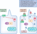
Figure 141.1
The sonic hedgehog (Shh) pathway in the pathogenesis of basal cell carcinoma.
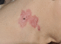
Figure 141.5
Superficial basal cell carcinoma.
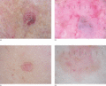
Figure 141.9
Dermoscopic images of basal cell carcinoma. (a) Nodular basal cell carcinoma. (b) Corresponding dermoscopic image showing white and grey‐brown structu...
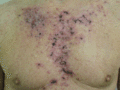
Figure 141.13
Skin reaction to topical imiquimod therapy for superficial basal cell carcinoma.
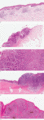
Figure 141.2
Histopathological patterns of basal cell carcinoma. (a) Superficial. Skin with small islands and downgrowths of basaloid cells arising from multiple p...
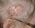
Figure 141.6
Morphoeic basal cell carcinoma.
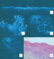
Figure 141.10
Nodular basal cell carcinoma (BCC). (a) H&E stained histological section shows actinic changes in overlying epidermis, islands of basaloid cells with ...
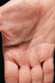
Figure 141.14
Palmar pits in a patient with Gorlin syndrome.
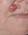
Figure 141.3
Nodular basal cell carcinoma.
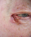
Figure 141.7
Ulcerated basal cell carcinoma.
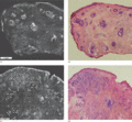
Figure 141.11
Fluorescent confocal submosaics (a,c) and the corresponding H&E stained Mohs frozen sections (b,d) at 4× magnification. The submosaic and frozen secti...
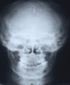
Figure 141.15
Skull X‐ray with calcified falx cerebri and jaw cyst.
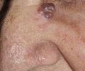
Figure 141.4
Pigmented basal cell carcinoma.
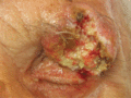
Figure 141.8
Advanced basal cell carcinoma.
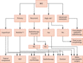
Figure 141.12
An algorithm for the treatment of basal cell carcinomas (BCC). *High‐risk BCCs include morphoeic lesions/infiltrative and micronodular growth pattern/...
