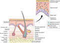
Figure 02.1
The skin and its appendages.
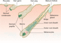
Figure 02.5
Embryonic stages of hair follicle morphogenesis.
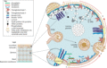
Figure 02.9
Assembly of the epidermal cornified cell envelope. In response to increasing intracellular calcium, an internal scaffold of desmosomal proteins is mad...
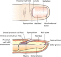
Figure 02.13
Anatomy and structure of the human nail.
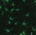
Figure 02.17
Dendritic appearance of epidermal Langerhans cells. Exposure to antigen provokes an increased movement of Langerhans cells as well as direct cell–cell...
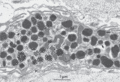
Figure 02.21
Part of a human skin mast cell showing characteristic granules, some with scroll‐like profiles (S). Arrows indicate perigranular membrane; L, lipid dr...
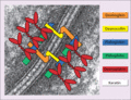
Figure 02.25
Macromolecular composition of desmosomes linking adjacent keratinocytes. Cells are connected via transmembranous cadherin glycoproteins (desmogleins a...
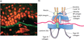
Figure 02.29
NaCl‐induced separation between the epidermis and dermis and antigen mapping within the cutaneous basement membrane. (a) The dermal–epidermal basement...
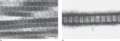
Figure 02.33
Demonstration of periodicity in collagen fibres of 640 Å. (a) Collagen fibrils in the reticular dermis show a characteristic banding pattern after sta...
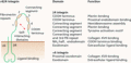
Figure 02.37
Molecular composition, domain organization and functions of the α6β4 integrin, the main keratinocyte integrin in hemidesmosomes. This integrin is impo...
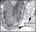
Figure 02.41
Transmission electron microscopy of the dermal–epidermal junction revealing wheatsheaf‐shaped anchoring fibrils beneath the lamina densa. These fibril...
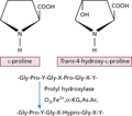
Figure 02.45
Enzymatic hydroxylation of prolyl residues in the Y‐position of the repeating Gly‐X‐Y amino acid sequence to form hydroxyproline, an amino acid charac...
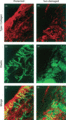
Figure 02.49
Immunofluorescence staining of type I collagen (a,d) and the elastic fibre network (b,e) in the dermis of human skin visualized by confocal laser scan...
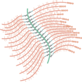
Figure 02.53
Prototypic proteoglycan in which the central core (green) is hyaluronic and the link proteins are represented by S‐shapes, joining the protein side ch...
Figure 02.57
Transmission electron microscopy of an activated dermal fibroblast (F) in a healing wound. Note the prominent rough endoplasmic reticulum in the cytop...
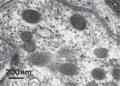
Figure 02.61
High magnification view of Weibel–Palade bodies revealing tubular profiles in cross‐section. (Courtesy of Professor R. A. J. Eady, St John's Institut...
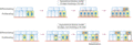
Figure 02.65
Possible mechanisms for the proliferative potential of stem cells (SC) in the basal keratinocyte layer. In the symmetrical division model, two stem ce...
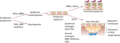
Figure 02.2
Embryonic development of the skin depends on specific signalling molecules. Relative stimulation or inhibition by these signalling molecules also dete...
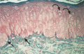
Figure 02.6
Photomicrograph of a 1 μm‐thick plastic section of normal human skin. The tissue was fixed with half‐strength Karnovsky medium and embedded in Epon. T...
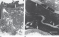
Figure 02.10
Electron micrograph showing the location of epidermal lipids by ruthenium oxide staining. (a) Extrusion of lamellar body lipids or sheets can be seen ...
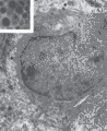
Figure 02.14
Merkel cell in human epidermis. The dermis (d) with collagen fibres is seen in the lower part of the picture; b, basement membrane; de, desmosomes mak...
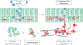
Figure 02.18
When exposed to foreign antigen, the activity of resting Langerhans cells increases and the cells mature. Antigen is then processed and transported to...
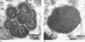
Figure 02.22
High‐magnification views of dermal mast cell granules. (a) Typical scroll‐like configuration of lamellae, some of which show a cross‐banding of regula...
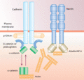
Figure 02.26
Macromolecular composition of an adherens junction in keratinocytes. There are two main components, nectin–afadin and the classic cadherin–catenin com...
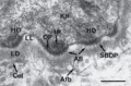
Figure 02.30
Transmission electron microscopy of the dermal–epidermal junction in human skin recognizing hemidesmosomes (HD), anchoring filaments (Afl) and anchori...
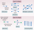
Figure 02.34
Assembly of type IV and type VII collagen molecules into supramolecular structures. The red boxes represent intermolecular disulphide bonds.
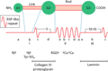
Figure 02.38
Model of nidogen, containing subdomains with predicted binding activities to type IV collagen, proteoglycans and laminin 1. EF and Ca refer to putativ...
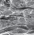
Figure 02.42
Transmission electron micrograph of a section of dermis from the human forearm showing bundles of collagen fibres, both in transverse and longitudinal...
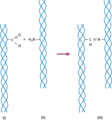
Figure 02.46
Formation of intermolecular cross‐links between individual collagen molecules. The cross‐linking is initiated by the conversion of lysine or a hydroxy...
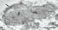
Figure 02.50
Transmission electron microscopy of an elastic fibre in the reticular dermis. The central electron‐pale core consists of elastin (E), while the electr...
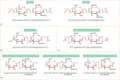
Figure 02.54
Glycosaminoglycan molecules that comprise the carbohydrate polymer side chains of proteoglycan molecules, including (a) heparin and heparan, (b) hyalu...
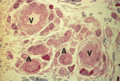
Figure 02.58
Histology of microvessels in the reticular dermis. Arterioles (A) can be distinguished from venules (V) by the presence of elastic lamina, which stain...
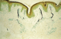
Figure 02.62
Histochemical detection of alkaline phosphatase activity indicating the presence of arterial microvessels in the superficial dermis. Original magnific...
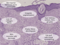
Figure 02.66
Structural and functional changes associated with skin ageing.
Figure 02.3
Scanning electron micrograph of an 85–110‐day (estimated gestation age) human embryo. Single globular blebs project from the periderm cells. (Courtes...
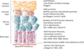
Figure 02.7
The process of epidermal differentiation is associated with the expression of different structures, macromolecules, transcription factors and other si...
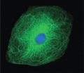
Figure 02.11
Structural organization of the keratin filament network within a keratinocyte. (Courtesy of Professor W. H. I. McLean, University of Dundee, UK.)
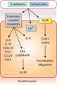
Figure 02.15
As part of the innate immune defence system, antimicrobial peptides can stimulate G‐protein‐coupled receptors to induce cytokine and chemokine release...
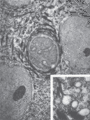
Figure 02.19
Langerhans cell (L) with its characteristically indented nucleus, situated between keratinocytes. The inset shows Langerhans cell granules with racque...
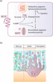
Figure 02.23
(a) Activation of the melanocortin 1 receptor (MC1R) promotes the synthesis of eumelanin at the expense of phaeomelanin. Oxidation of tyrosine by tyro...
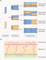
Figure 02.27
Formation and structure of gap junctions in human skin. (a) In the Golgi network six connexin subunits assemble to form a connexon. The connexon is th...
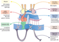
Figure 02.31
Gene/protein systems within the cutaneous basement membrane zone that can harbour mutations and result in blistering of the skin in different forms of...
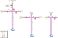
Figure 02.35
Different isoforms and domain organizations of laminin, each consisting of three distinct subunit polypeptides, α‐, β‐ and γ‐chains. The LE modules ar...
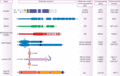
Figure 02.39
Structure and domain organization of the major protein components at the cutaneous basement membrane zone, with their molecular weights and chromosoma...
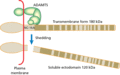
Figure 02.43
Type XVII collagen, a transmembrane protein in type 2 orientation. Note that the ectodomain traversing the lamina lucida contains 15 distinct triple h...
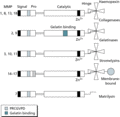
Figure 02.47
Structural organization of various matrix metalloproteinases (MMPs), divided into different subclasses. The signal, propeptide, active catalytic hinge...
Figure 02.51
Assembly and cross‐linking of elastic fibres. Newly synthesized elastin precursor polypeptides, tropoelastins, with alternating hydrophobic and cross‐...
Figure 02.55
Core protein aggrecan is joined by link proteins to hyluronate, with keratan sulphate (KS, blue) and chondroitin sulphate (CS, purple) side chains. aa...
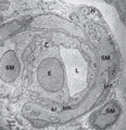
Figure 02.59
Transmission electron microscopy of a cross‐section through a small arteriole in the skin. Note the relatively smooth surface of the endothelial cell ...
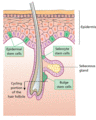
Figure 02.63
Epithelial stem cells are found within the interfollicular epidermis, the base of sebaceous glands and in the bulge area of hair follicles.
Figure 02.4
Electron micrograph of the full‐thickness epidermis from the back of a 14‐week human fetus. The periderm cells are full of glycogen (g) and have micro...
Figure 02.8
Electron micrograph showing details of the upper part of the epidermis including the stratum corneum (SC), stratum granulosum (SG) and most superficia...
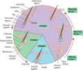
Figure 02.12
There are three components to the hair cycle: anagen (where new hair forms and grows), followed by catagen (regressing phase) and telogen (resting pha...
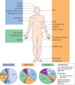
Figure 02.16
The skin microbiome contains numerous bacteria that are variably present in different body regions. (Adapted from Chen and Tsao 2013 [ ].)
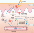
Figure 02.20
Immune surveillance in normal skin is carried out by an array of skin‐based dendritic cells, macrophages and resident T cells. iNOS, inducible nitric ...
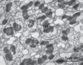
Figure 02.24
Electron micrograph of desmosomes in the spinous layer. These intercellular junctions are closely associated with tonofilaments (tf), many of which, i...
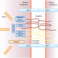
Figure 02.28
Structural composition of a tight junction in human skin. There are three transmembranous families of proteins, the junctional adhesion molecules (JAM...
Figure 02.32
Immunofluorescence staining of the dermis and cutaneous basement membrane zone with an antibody for type IV collagen. Note positive staining at the de...
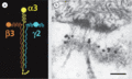
Figure 02.36
(a) Laminin 332 expressed in the cutaneous basement membrane zone. (b) Immunogold electron microscopy using an antibody to the γ2 chain of laminin 332...
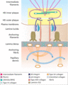
Figure 02.40
Molecular interactions of the major components of the cutaneous basement membrane zone. The individual components are identified in the colour key and...
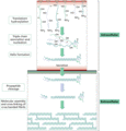
Figure 02.44
Steps in the intracellular biosynthesis of triple helical type I procollagen, its secretion into the extracellular space, and assembly and cross‐linki...
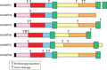
Figure 02.48
Main organization of various ADAMTS family metalloproteinases. The catalytic domain is shown in red, while other domains include the thrombospondin ty...

Figure 02.52
Elastic fibres cross‐linked by desmosines (red). In the relaxed state, the fibres assume coiled‐coil conformations. When the fibres are stretched and ...
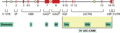
Figure 02.56
Human versican gene: intron–exon organization (top) and deduced functional domains of the encoded protein. CRP, complement regulatory protein; EGF, ep...
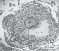
Figure 02.60
Transmission electron microscopy of a transverse section through a venule in the skin. The surface of the endothelial cells (E) in the lumen (L) is mo...
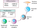
Figure 02.64
Epidermal stem cell proliferation is regulated positively by β1 integrin and transforming growth factor α (TGF‐α) and negatively (–) by TGF‐β signalli...
