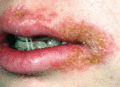
Figure 26.1
Staphylococcal impetigo. (Courtesy of King's College Hospital Dermatology Department, London, UK.)
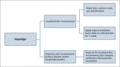
Figure 26.5
Management of impetigo.
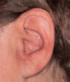
Figure 26.9
Poststreptococcal lymphoedema of the pinna; this patient had frequent recurrences of cellulitis requiring long‐term penicillin.
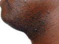
Figure 26.13
Pseudofolliculitis.
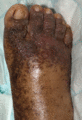
Figure 26.17
Indolent blistering associated with toxic shock syndrome.
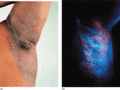
Figure 26.21
(a) Erythrasma in the axilla. (Courtesy of St John's Institute of Dermatology, London, UK.) (b) Fluorescence with Wood's light. (Courtesy of King's C...
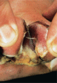
Figure 26.25
Pseudomonas infection of the foot. (Courtesy of St John's Institute of Dermatology, King's College London, UK.)
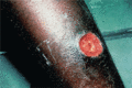
Figure 26.29
Tropical ulcer. (Courtesy of St John's Institute of Dermatology, London, UK.)
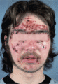
Figure 26.2
Streptococcal (group A) pyoderma.
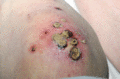
Figure 26.6
Ecthyma. (Courtesy of King's College Hospital Dermatology Department, London, UK.)
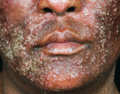
Figure 26.10
Acute folliculitis on the face. (Courtesy of King's College Hospital Dermatology Department, London, UK.)
Figure 26.14
Staphylococcal scalded skin syndrome in a child. (Courtesy of King's College Hospital Dermatology Department, London, UK.)
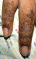
Figure 26.18
Retiform purpura in toxic shock syndrome.
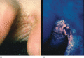
Figure 26.22
(a,b) Erythrasma of the toe cleft. (b) Fluorescence with Wood's light. (Courtesy of King's College Hospital Dermatology Department, London, UK.)
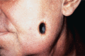
Figure 26.26
Ecthyma gangrenosum. (Courtesy of Dr G. Scott, University College Hospital, London, UK.)
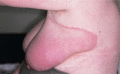
Figure 26.30
Erythema chronicum migrans. (Courtesy of Dr A. S. Highet, York District Hospital, York, UK.)
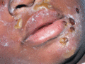
Figure 26.3
Bullous impetigo. (Courtesy of King's College Hospital Dermatology Department, London, UK.)
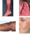
Figure 26.7
Cellulitis/erysipelas. (a) Lower leg. (b) Bullous cellulitis of the leg. (Courtesy of King's College Hospital Dermatology Department, London, UK.) (...
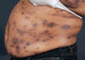
Figure 26.11
Staphylococcus aureus abscesses. (Courtesy of King's College Hospital Dermatology Department, London, UK.)
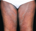
Figure 26.15
Staphylococcal scalded skin syndrome in an adult. (Courtesy of King's College Hospital Dermatology Department, London, UK.)
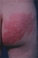
Figure 26.19
Recurrent toxin‐mediated perineal erythema.
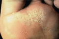
Figure 26.23
Pitted keratolysis. (Courtesy of St John's Institute of Dermatology, King's College London, UK.)
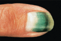
Figure 26.27
Pseudomonas infection of the nail. (Courtesy of St John's Institute of Dermatology, King's College London, UK.)
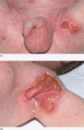
Figure 26.4
(a,b) Erosive bullous impetigo in a neonate. (Courtesy of King's College Hospital Dermatology Department, London, UK.)
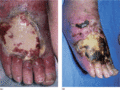
Figure 26.8
(a) Cellulitis with early dermal necrosis. (b) The same foot after 11 days; the dermis is forming a black eschar, which eventually sloughed off; the r...
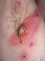
Figure 26.12
Panton–Valentine leukocidin multiple necrotic recurrent abscesses. (Courtesy of King's College Hospital Dermatology Department, London, UK.)
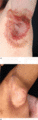
Figure 26.16
(a) Localized staphyloccocal scalded skin syndrome (SSSS). (b) Localized SSSS healing with wrinkling desquamation and hyperpigmentation. (Courtesy of...
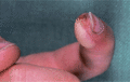
Figure 26.20
Blistering distal dactylitis.
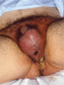
Figure 26.24
Fournier necrotizing fasciitis of the groin. (Courtesy of King's College Hospital Dermatology Department, London, UK.)
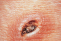
Figure 26.28
Pasteurella multocida infection. (Courtesy of St John's Institute of Dermatology, London, UK.)
