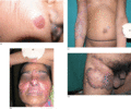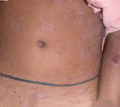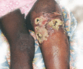
Figure 27.1
Warty tuberculosis. (a) Epidermis showing hyperkeratosis and acanthosis, with acute inflammation and abscess formation in the upper dermis. Ill‐define...

Figure 27.5
Mycobacterium bovis BCG infection of the glans penis, showing an infiltrated red plaque containing small, deep‐seated, yellow papules. (Courtesy of ...

Figure 27.9
Tuberculous gumma (metastatic cold abscess) in a patient with a pleural effusion. (Courtesy of Dr V. Ramesh, SJ Hospital and VM Medical College, New ...

Figure 27.13
Sporotrichoid spread of warty tuberculosis. (Courtesy of Dr V. Ramesh, SJ Hospital and VM Medical College, New Delhi, India, and the Editor of Derma...

Figure 27.17
Tumour‐like form of lupus vulgaris on the ear lobe and face. (Courtesy of Dr V. Ramesh, SJ Hospital and VM Medical College, New Delhi, India, and the...

Figure 27.21
Lichen scrofulosorum of the trunk showing subtle skin‐coloured papules. (Courtesy of Dr Antonio Torrelo, Hospital del Niño Jesus, Madrid, Spain.)

Figure 27.25
Papulonecrotic tuberculid of the legs. (Courtesy of Professor J. Aboobaker, University of Natal, Durban, South Africa.)

Figure 27.29
Fish infected with Mycobacterium marinum . (Courtesy of Professor J. A. A. Hunter, Royal Infirmary, Edinburgh, UK.)

Figure 27.33
Mycobacterium avium complex infection. (a) Presenting as an erythematous nodule with central ulceration on the extensor aspect of the forearm in an i...

Figure 27.37
Mycobacterium chelonae infection of the lower limb. (Courtesy of Dr R. D. Ead, Hope Hospital, Manchester, UK.)

Figure 27.2
Lupus vulgaris. Extensive caseous granulomatous inflammation in the deep dermis in lupus vulgaris (H&E). (Courtesy of Dr M. Bamford and Dr A. Fletche...

Figure 27.6
Scrofuloderma. (a) Associated with tuberculosis of the axillary glands occurring in a 74‐year‐old white man prior to antituberculous therapy. (b) Asso...

Figure 27.10
Multiple metastatic abscesses (gummas) on the left arm. (Courtesy of Dr V. Ramesh, SJ Hospital and VM Medical College, New Delhi, India, and the Edit...

Figure 27.14
Lupus vulgaris. (a) A solitary plaque on the left cheek. (b) Lesions of the face resembling discoid lupus erythematosis. Note the strong tuberculin re...

Figure 27.18
Perianal lupus (a) before and (b) after treatment. (Courtesy of Dr Julia Rhodes, Department of Dermatology, Royal Perth Hospital, Australia, and the ...

Figure 27.22
Lichen scrofulosorum showing annular and plaque‐like lesions with strong tuberculin reaction. (Courtesy of Dr V. Ramesh, SJ Hospital and VM Medical C...

Figure 27.26
Erythema induratum of the legs.

Figure 27.30
Mycobacterium marinum infection showing sporotrichoid spread from the hand to the forearm in an aquarist. (Courtesy of Dr I. H. Coulson, Burnley Gen...

Figure 27.34
Mycobacterium haemophilum infection producing ulcerated nodules on the left knee and shin, with a non‐ulcerated nodule on the medial aspect of the kn...

Figure 27.3
Plaque of lupus vulgaris measuring 50 × 30 mm at the site of a previous BCG vaccination. (Courtesy of Dr S.L. Walker, Faculty of Infectious and Tropi...

Figure 27.7
(a) Ulcerative form of scrofuloderma in an Ethiopian child secondary to tuberculous osteomyelitis of the skull. (b) The same child after several month...

Figure 27.11
Warty tuberculosis with strong tuberculin reactions. (a) The right ring and middle fingers. (b) Sole of the left foot. (Courtesy of Dr V. Ramesh, SJ ...

Figure 27.15
Nasal deformity as a late sequela of lupus vulgaris. (Courtesy of Dr V. Ramesh, SJ Hospital and VM Medical College, New Delhi, India, and the Editor ...

Figure 27.19
Extensive BCG‐induced lupus vulgaris in a child complicated by squamous cell carcinoma. (Courtesy of Dr Binod Kr Thakur and Dr Shikha Verma, North Ea...

Figure 27.23
Lichen scrofulosorum on the forehead. (Courtesey of Dr Yogesh S. Marfatia, Department of Skin‐VD, Medical College and SSG Hospital, Vadodara, Gujarat,...

Figure 27.27
Acneform‐like lesions with scarring on the face and ears. Tuberculoid histology with acid‐fast bacilli is seen demonstrating a true lupus miliaris dis...

Figure 27.31
Mycobacterium ulcerans in an 8‐year‐old child. (Courtesy of Professor F. Portaels, Institute of Tropical Medicine, Antwerp, Belgium.)

Figure 27.35
Mycobacterium abscessus infection. (a) Cellular infiltrate in the mid and deep dermis. (b) A heavy infiltrate of neutrophils and nuclear dust in the ...

Figure 27.4
Atypical papular tuberculid following BCG vaccination. (Courtesy of Dr J. Muto, National Defence Medical College, Namiki, Tororozawa, Saitama, Japan....

Figure 27.8
Periorificial tuberculosis. (a) Crusty erosions are evident on the gingival surface and mucosal surface of the lip. (Courtesy of Dr F. Nachbar, Ludwi...

Figure 27.12
Warty tuberculosis of the right thigh. (Courtesy of Dr V. Ramesh, SJ Hospital and VM Medical College, New Delhi, India, and the Editor of Paediatric...

Figure 27.16
Vegetating lupus vulgaris on the nose. (Courtesy of Dr J. E. Bothwell, Barnsley District General Hospital, Barnsley, UK.)

Figure 27.20
A basal cell carcinoma arising in an old area of lupus vulgaris.

Figure 27.24
Lichenoid variety of lichen scrofulosorum. (Courtesy of Dr Luis Requena, Fundación Jiménez Diaz, Universidad Autónoma, Madrid, and the Editor of the ...

Figure 27.28
Granulomatous mastitis due to tuberculosis. (Courtesy of Dr F. G. Bravo, Instituto de Medicina Tropical Alexander von Humboldt, Lima, Peru.)

Figure 27.32
Extensive Mycobacterium ulcerans of the elbow in a child. (Courtesy of Dr P. L. A. Niemel, Surinam.)

Figure 27.36
Mycobacterium abscessus infection causing abscesses. (Courtesy of Dr A. G. Smith, North Staffordshire Hospital, Stoke‐on‐Trent, UK, and the Editor o...

