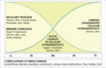
Figure 28.1
Mechanisms of damage in leprosy, and tissues affected. Mechanisms under the broken line are characteristic of disease near the lepromatous end of the ...
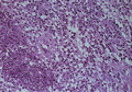
Figure 28.5
Erythema nodosum leprosum. Medium‐power view (H&E) showing foamy macrophages with infiltrating polymorphs. There is also a swollen small artery in the...
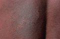
Figure 28.9
Tuberculoid or borderline tuberculoid leprosy. Back of a Nigerian child showing well‐defined hypopigmented macule with altered skin texture. The lesio...
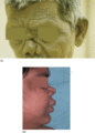
Figure 28.13
Lepromatous leprosy. (a) Man with nasal collapse and forehead wrinkling. (b) Woman with nasal collapse.

Figure 28.17
Borderline tuberculoid leprosy. Back of Nigerian man showing large well‐defined scaly macules with some marginal elevation. Extensive disease, such as...
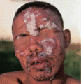
Figure 28.21
Type 1 reaction: borderline leprosy in an Ethiopian man. Existing lesions become acutely inflamed, scale and threaten ulceration. Many small new lesio...
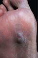
Figure 28.25
Necrosis blister in an anaesthetic foot. Necrotic material has tracked to soft skin from the site of trauma, beneath the metatarsal heads.
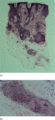
Figure 28.2
(a) Low‐power view (H&E) showing tuberculoid granulomas around nerve and skin appendages in mid‐dermis and a swollen, deep dermal nerve. There is no e...
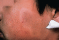
Figure 28.6
Indeterminate leprosy. Face of a Nepali child showing vague hypopigmented patch with some central healing. Note the mark of a recent slit‐skin smear.

Figure 28.10
Lepromatous leprosy (borderline lepromatous/lepromatous. Back of a Bangladeshi boy showing numerous, often confluent hypopigmented macules, with relat...
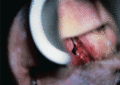
Figure 28.14
Lepromatous leprosy. Examination via a nasal speculum shows a pale septal nodule and bleeding. Such lesions in untreated lepromatous patients constitu...
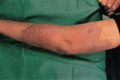
Figure 28.18
Borderline tuberculoid leprosy. Arm of an Indian woman. A large scaly macule is developing secondary ichthyotic change.
Figure 28.22
Type 2 reaction in lepromatous leprosy in a Nigerian man: erythema nodosum leprosum. Several of the reaction nodules have broken down, releasing pus.
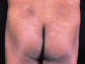
Figure 28.26
Borderline leprosy (BL). Borderline leprosy upgrading to borderline tuberculoid (BT). Buttocks of an Indian man who ‘relapsed’ 3 years after completin...
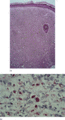
Figure 28.3
(a) Lepromatous leprosy. Medium‐power view (H&E) showing thin epidermis, a clear subepidermal zone and a dense, uniform macrophage infiltrate in the d...
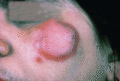
Figure 28.7
Tuberculoid leprosy. Face of Pakistani woman showing erythematous plaque with a well‐defined active edge, and a small satellite lesion. On the face, s...
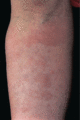
Figure 28.11
Lepromatous leprosy. Forearm of an English man showing erythematous macules and infiltration.
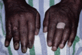
Figure 28.15
Lepromatous leprosy. Hands of an Indian man, showing swollen fingers due to leprous dactylitis and one crooked finger due to a pathological fracture. ...
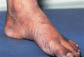
Figure 28.19
Borderline leprosy. Foot of a Bangladeshi child showing enlargement of posterior tibial and anterior tibial nerves.
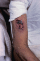
Figure 28.23
Type 2 reaction in lepromatous leprosy in a Bangladeshi man, showing severe necrosis and ulceration. Erythema nodosum leprosum tends to be more severe...
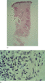
Figure 28.4
(a) Indeterminate leprosy. Low‐power view (H&E) showing minimal perineurovascular inflammation in the mid‐dermis. (b) High‐power view, Wade–Fite stain...
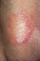
Figure 28.8
Tuberculoid or borderline tuberculoid leprosy. Upper arm of Indian man, showing typical dry, hairless, hypopigmented plaque with scaly, erythematous e...
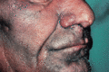
Figure 28.12
Lepromatous leprosy. Face of a man showing diffuse infiltration of the skin and appearance of nodules on the nose and lip.
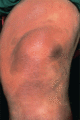
Figure 28.16
Borderline leprosy. Knee of Saudi‐Arabian woman, showing classical annular lesions with well‐defined centres. Slit‐skin smears show acid‐fast bacilli....
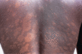
Figure 28.20
Borderline leprosy (BL). Borderline tuberculoid (BT) downgrading to BL. Back of a Nigerian woman, showing typical well‐defined hypopigmented macules o...
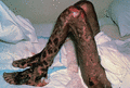
Figure 28.24
Lucio phenomenon in a Mexican man. The severe recurrent ulceration due to deep subcutaneous vasculitis may be fatal. (Courtesy of Dr J. Keystone, the...
