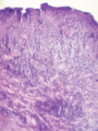
Figure 48.1
Abundant mixed inflammatory infiltrate dominated by neutrophils in an oral ulcer of Adamantiades–Behçet disease.
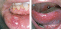
Figure 48.2
Single (a) and multiple (b) oral aphthous ulcers. (Part (a) from Altenburg et al . 2006 [ ]. Reproduced with permission of John Wiley & Sons.)
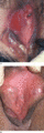
Figure 48.3
Genital ulcer (a) healing with a demarcated flat scar (b).
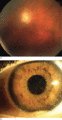
Figure 48.4
(a) Posterior uveitis. (b) Hypopyoniritis. (From Altenburg et al . 2006 [ ]. Reproduced with permission of John Wiley & Sons.)
