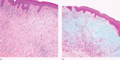
Figure 59.1
(a) Scleromyxoedema. The typical triad of microscopic features with diffuse dermal mucin deposition, fibroblast proliferation and fibrotic collagen. (...
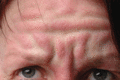
Figure 59.5
Scleromyxoedema. Deep longitudinal erythematous folding on the forehead (leonine face). (Courtesy of D. Metze, MD, Munster, Germany.)
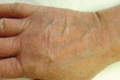
Figure 59.9
Acral persistent papular mucinosis. Multiple skin‐coloured papules on the dorsal aspect of the hand.
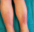
Figure 59.13
Pretibial myxoedema with diffuse non‐pitting oedema and plaque‐like lesions on the legs.
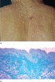
Figure 59.17
(a) Papular and nodular mucinosis in connective tissue disease. (b) Abundant mucin deposition throughout the reticular dermis (Alcian blue stain).
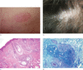
Figure 59.21
(a) Pinkus follicular mucinosis. A sharply demarcated erythematous plaque with follicular prominence. (b) Alopecic plaque in follicular mucinosis. (c)...
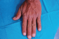
Figure 59.2
Scleromyxoedema. Widespread eruption of closely spaced papules on the back of the hand.
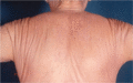
Figure 59.6
Scleromyxoedema. Deep furrowing on the shoulders and back (Shar‐Pei sign).
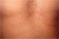
Figure 59.10
Discrete papular lichen myxoedematosus. Mucinous skin‐coloured papules on the trunk.
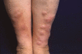
Figure 59.14
Pretibial myxoedema of nodular type.
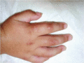
Figure 59.18
Self‐healing cutaneous mucinosis. Mucinous subcutaneous nodules and papules on the periarticular areas of the hand in a child.
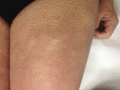
Figure 59.3
Scleromyxoedema. Papules on the thigh.
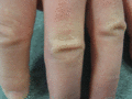
Figure 59.7
Scleromyxoedema. ‘Doughnut sign’ on the proximal interphalangeal joints.
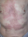
Figure 59.11
Reticular erythematous mucinosis in the midline of the chest and abdomen.
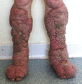
Figure 59.15
Elephantiasic pretibial myxoedema. (Courtesy of B. Cribier, MD, Strasbourg, France.)
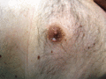
Figure 59.19
Cutaneous focal mucinosis. A solitary whitish papule with cystic appearance.
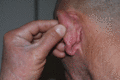
Figure 59.4
Scleromyxoedema. Papules behind the ear.
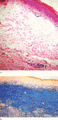
Figure 59.8
(a) Microscopic features of acral persistent papular mucinosis. Focal mucin accumulation in the upper dermis sparing a grenz zone without fibroblast p...
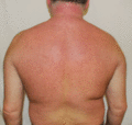
Figure 59.12
Scleredema in a diabetic patient with firm non‐pitting oedema and induration on the upper back, neck and shoulders on erythematous background.
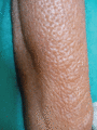
Figure 59.16
Localized myxoedema on the preradial area. Note the orange peel appearance. (Courtesy of S. Verma, MD, Vadodara, Gujarat, India.)
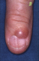
Figure 59.20
Digital myxoid cyst. A translucent dome‐shaped nodule on the distal interphalangeal joint of the finger.
