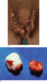
Figure 61.1
(a) Calcinosis scrotalis. (b) Solitary nodules after removal.
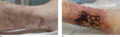
Figure 61.5
(a) Typical livedo in early calciphylaxis on the lower leg. (b) Progression to ulceration with surrounding livedo.
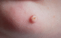
Figure 61.2
Idiopathic calcinosis cutis on the chin of a child.
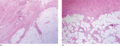
Figure 61.6
(a) Calcification of two arterioles. (b) Calcification of small vessels in a background of fat necrosis. (Courtesy of Dr J. Fitzgibbon, Cork Universi...
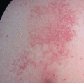
Figure 61.3
Miliary calcinosis cutis.
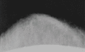
Figure 61.7
Soft tissue radiography showing calcification of vessels of the abdominal wall. (Reproduced with permission from Hackett BC et al. Calciphylaxis in a...
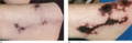
Figure 61.4
Calciphylaxis with painful purpuric lesions on the thigh in a 53‐year‐old diabetic patient with renal failure. (a) At presentation. (b) Progressing to...
