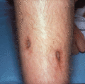
Figure 64.1
Diabetic dermopathy on both shins.
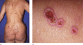
Figure 64.5
(a) Perforating collagenosis on the back of a 64‐year‐old woman with diabetic retinopathy. (b) Close‐up view.
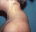
Figure 64.9
Scleredema diabeticorum with a ‘buffalo hump’ in a young woman with diabetes.
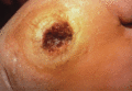
Figure 64.2
Diabetic foot with neurotrophic ulceration and necrosis (‘mal perforans’).
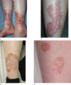
Figure 64.6
(a–d) Necrobiosis lipoidica with ulcerations on the shins at various stages of evolution.
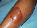
Figure 64.10
A large diabetic bulla on the shin of a patient with diabetes.

Figure 64.3
Velvety warty hyperpigmentation on the front (a) and back (b) of the neck in acanthosis nigricans. (Courtesy of Dr Robin A. C. Graham‐Brown, Leiceste...
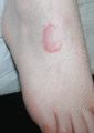
Figure 64.7
Granuloma annulare of the foot.
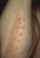
Figure 64.4
Eruptive xanthomas in a diabetic patient with hyperlipidaemia.
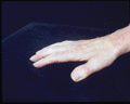
Figure 64.8
Cheirarthropathy of the hands. (Courtesy of Dr Robin A. C. Graham‐Brown, Leicester Royal Infirmary.)
