
Figure 81.1
Facial features of an 8‐year‐old boy with Hunter syndrome (MPS II).
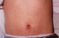
Figure 81.5
Angiokeratoma corporis diffusum around the umbilicus in a man with Fabry disease.
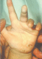
Figure 81.9
Hyperkeratotic lesion on the palm of a child with tyrosinaemia type 2.(From Sarafoglou et al . 2009 [ ]. Reproduced with permission of McGraw Hill E...
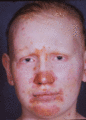
Figure 81.13
Acrodermatitis enteropathica with typical eczematous skin lesions. The vermilion area is free, and the hair growth poor and sparse.
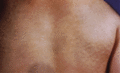
Figure 81.2
‘Pebbling’ of the skin and extensive Mongolian blue spot on the back of a boy with Hunter syndrome (MPS II). (From Wraith 2006 [ ]. Reproduced with p...
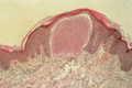
Figure 81.6
Angiokeratoma corporis diffusum showing dilated blood‐filled vessels in the papillary dermis.
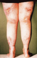
Figure 81.10
Multiple ulcers with an unusual distribution associated with prolidase deficiency.

Figure 81.14
Infant with Menkes disease. (Courtesy of Dr A. Chakrapani, Birmingham Children's Hospital, Birmingham, UK.)
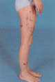
Figure 81.3
Angiokeratoma corporis diffusum on the limbs of a 7‐year‐old girl with fucosidosis.
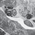
Figure 81.7
Angiokeratoma corporis diffusum. Electron‐dense cytoplasmic inclusion bodies are present within the endothelial cells. (Courtesy of Dr P. H. McKee, K...
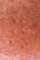
Figure 81.11
Pitted skin in prolidase deficiency. (Courtesy of Dr D. A. Burns, Leicester Royal Infirmary, Leicester, UK.)
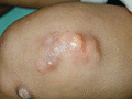
Figure 81.15
Calcinosis knee of a child with normophosphataemic familial tumoral calcinosis.
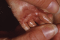
Figure 81.4
The hand of a ‘collodion baby’ with Gaucher disease type II.
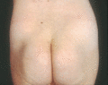
Figure 81.8
Fat pads over the iliac crests in a child with phosphomannomutase 2 deficiency. (Courtesy of Professor P. T. Clayton, Institute of Child Health, Lond...

Figure 81.12
Trichorrhexis nodosa in a 4‐year‐old with argininosuccinic aciduria. (Courtesy of Central Manchester University Hospitals NHS Foundation Trust, Manch...
