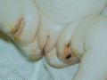
Figure 82.1
Ulcerated perineal region in an infant with severe combined immunodeficiency.
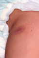
Figure 82.5
Cutaneous lesion in a child with severe combined immunodeficiency who had been immunized with the live bacille Calmette–Guérin (BCG) vaccine. On biops...
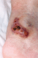
Figure 82.9
Vasculitis in a boy with X‐linked lymphoproliferative disease.
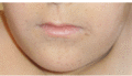
Figure 82.13
Characteristic perioral reticular hyperpigmentation in dyskeratosis congenita.
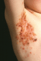
Figure 82.17
Childhood linear IgA disease in FAS‐deficient autoimmune lymphoproliferative syndrome.
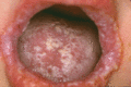
Figure 82.21
Oro‐pharyngeal mucocutaneous Candida infection in a patient with a gain‐of‐function STAT1 mutation.
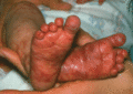
Figure 82.2
Maternofetal graft‐versus‐host disease in an infant with JAK3‐deficient severe combined immunodeficiency.
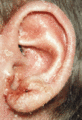
Figure 82.6
Severe eczema in a child with Wiskott–Aldrich syndrome.
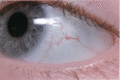
Figure 82.10
Bulbar telangiectasia in a patient with ataxia telangiectasia.
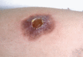
Figure 82.14
Cellulitis due to Pseudomonas infection in a patient with X‐linked agammaglobulinaemia.

Figure 82.18
Translucent papular lesions around the eyelids of a patient with chronic granulomatous disease.
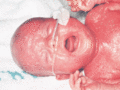
Figure 82.3
Omenn syndrome, with characteristic erythroderma and alopecia.
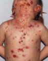
Figure 82.7
Severe molluscum contagiosum in a child with DOCK8 deficiency.
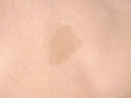
Figure 82.11
Hyperpigmented area on the back of a patient with Bloom syndrome.
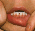
Figure 82.15
Partial albinism in Chediak–Higashi syndrome.
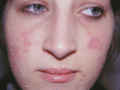
Figure 82.19
Malar erythematous photosensitive macular skin lesions in an X‐linked carrier of chronic granulomatous disease.
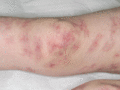
Figure 82.4
Inflammatory granulomatous skin lesions in a child with atypical severe combined immunodeficiency.
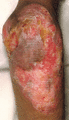
Figure 82.8
Sterile necrotizing granulomatous lesion on the knee of a patient with TAP1 deficiency.
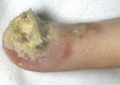
Figure 82.12
Extensive digital fungal infection in immunodeficiency, centromeric instability, facial dysmorphism syndrome.
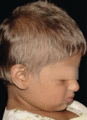
Figure 82.16
Patient with Griscelli syndrome demonstrating a silver sheen to the hair which has persisted post haematopoietic stem cell transplantation.
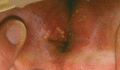
Figure 82.20
Erosive perianal ulcers in an infant with severe leukocyte adhesion deficiency type I.
