Figure 87.1
Acquired ichthyosis secondary to lymphoma.
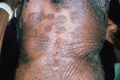
Figure 87.5
Pityriasis rotunda.
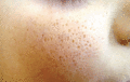
Figure 87.9
Atrophoderma vermiculatum the on the cheek of 10‐year‐old boy. (Source: http://www.ncbi.nlm.nih.gov/pmc/articles/PMC3163348/ from Apalla et al. J D...
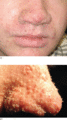
Figure 87.13
(a,b) Trichodysplasia spinulosa: multiple keratotic spicules on the nose of a heart transplant recipient. (Reproduced from PLOS Pathogens http://j...

Figure 87.17
Porokeratosis of Mibelli.
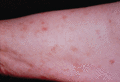
Figure 87.21
Disseminated superficial actinic porokeratosis: view of upper arm.
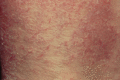
Figure 87.25
Xerosis cutis.
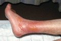
Figure 87.29
Diabetic bullae: recent development of extensive large bullae on the lower legs and feet in the absence of inflammation or oedema in a patient with lo...
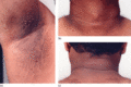
Figure 87.2
(a–c) Typical acanthosis nigricans in an obese 41‐year‐old man of South Asian descent with type II diabetes. Note associated striae and skin tags in t...
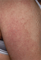
Figure 87.6
Keratosis pilaris on the extensor aspect of the upper arm.
Figure 87.10
Lichen spinulosus present for 18 months as an asymptomatic eruption on the back, shoulder and upper arm of an 8‐year‐old girl.
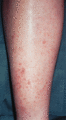
Figure 87.14
Flegel disease: multiple tiny thorn‐like keratotic papules on the skin of the lower leg.
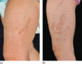
Figure 87.18
Linear porokeratosis: irregular linear and polygonal ‘Chinese character’ plaques developing in a blaschkoid distribution on the thigh, showing progres...
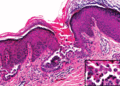
Figure 87.22
Transient acantholytic dermatosis: histopathological image of a papule demonstrating intraepidermal clefting and acantholytic cells (inset). (Reprodu...
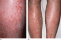
Figure 87.26
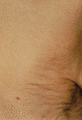
(a,b) Asteatotic eczema.
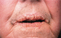
Figure 87.3
Malignant acanthosis nigricans: warty thickening of the oral margins in a patient with carcinoma of the breast.
Figure 87.7
Erythromelanosis follicularis faciei et colli in a young Asian man.
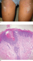
Figure 87.11
Keratosis circumscripta: coalescing hyperkeratotic papules on the thighs of 10‐year‐old African American girl (a); psoriasiform dermatitis with promin...
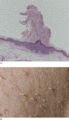
Figure 87.15
Multiple minute digitate keratoses: photomicrograph of hyperkeratotic spicule (a); close‐up view of spicules on the back (b). (From Caccetta et al . ...
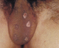
Figure 87.19
Genital porokeratosis: multiple lesions limited to the scrotum. (From Chen et al . 2006 [ ]. Reproduced with permission from the copyright holder Wi...
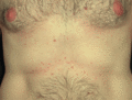
Figure 87.23
Transient acantholytic dermatosis: typical appearance on the abdomen. (Reproduced courtesy and with permission of Professor Luis Requena, Universidad...
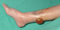
Figure 87.27
Acute oedema blisters in an elderly female: acute swelling of the lower limbs after withdrawal of diuretic therapy given for congestive heart failure ...
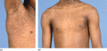
Figure 87.4
(a,b) Confluent and reticulate papillomatosis: an asymptomatic rash appeared 6 months earlier around the neck of this of 12‐year‐old boy before spread...
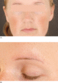
Figure 87.8
Keratosis pilaris atrophicans faciei (ulerythema ophryogenes): note well‐defined symmetrical erythema on the cheeks and above sparse residual eyebrow ...
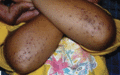
Figure 87.12
Phrynoderma: keratotic papules with intrafollicular plugging on extensor surfaces of the forearms of a 3‐year‐old Indian girl presenting with night bl...
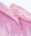
Figure 87.16
Cornoid lamella forming edge of a porokeratosis: a column of parakeratotic keratinocytes can be seen arising from invagination of the underlying epide...
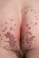
Figure 87.20
Perianal porokeratosis. (Courtesy of Dr P. Laws, Chapel Allerton Hospital, Leeds, UK.)
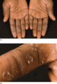
Figure 87.24
Keratolysis exfoliativa: view of the palms (a) with close‐up of right index finger (b).
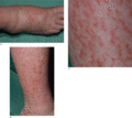
Figure 87.28
(a–c) Eczéma craquelé following acute onset of oedema due to congestive heart failure.
