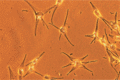
Figure 88.1
Melanocytes in culture.
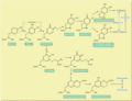
Figure 88.5
A simplified overview of the major metabolic pathways in the synthesis of melanins and trichochromes. (From Prota 1988 [ ], with permission from the c...
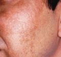
Figure 88.9
Melasma in an adult male from the Indian subcontinent.
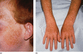
Figure 88.13
(a,b) Freckles in an 11‐year‐old boy.
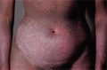
Figure 88.17
Generalized pigmentation in a woman aged 33 years with systemic sclerosis.
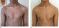
Figure 88.21
Vitamin B 12 deficiency: (a) generalized mottled hypermelanosis as presenting feature of pernicious anaemia in a 16‐year‐old boy with a 4‐year histor...
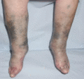
Figure 88.25
Minocycline pigmentation: marked dyspigmentation of the lower legs resulting from minocycline therapy commenced 2 years earlier as adjunctive therapy ...
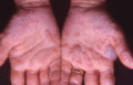
Figure 88.29
Phytophotodermatitis: acute irregular blisters across the palms after grasping giant hogweed.
Figure 88.33
Algorithm for the differential diagnosis of hypomelanosis.
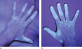
Figure 88.37
(a,b) Typical distribution of vitiligo on dorsum of the hand seen under Wood's light.

Figure 88.41
Hypochromic vitiligo affecting the back. (From Ezzedine et al . 2015 [ ] reproduced with permission from the copyright holder Wiley.)
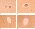
Figure 88.45
Halo naevi in different stages of evolution concurrently in a 13‐year‐old girl: (a) early depigmentation; (b) established halo naevus; (c) faint pink ...
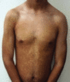
Figure 88.49
Progressive macular hypomelanosis in an 18‐year‐old man. (Reproduced with permission from the copyright holder Springer Publishing Company, from Rely...
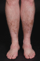
Figure 88.53
Haemosiderin staining on the shins of a 41‐year‐old rugby football player resulting from repeated minor trauma.
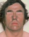
Figure 88.57
Occupational argyria.
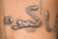
Figure 88.61
Keloid reaction to decorative tattoo: flattened areas have responded to the injection of triamcinolone.
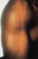
Figure 88.2
Voigt–Futcher lines. (Courtesy of the late Dr R. R. M. Harman, Bristol Royal Infirmary, Bristol, UK.)
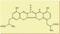
Figure 88.6
Structure of trichochrome B, one of six trichochromes so far identified.
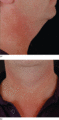
Figure 88.10
(a,b) Poikiloderma of Civatte: showing submental and submandibular sparing on the neck of a 43‐year‐old man.
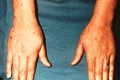
Figure 88.14
Eruptive lentiginosis: 4‐month history of widespread eruption of lentigines over the neck, trunk and limbs in a healthy 49‐year‐old female. Note lenti...
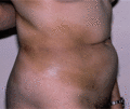
Figure 88.18
Morphoea. Hyperpigmentation was the presenting symptom.
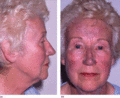
Figure 88.22
(a,b) Amiodarone pigmentation after 5 years of therapy: note slaty‐blue dyspigmentation of the forehead, nose, cheeks and earlobe.
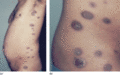
Figure 88.26
(a,b) Pigmented fixed drug eruption: extensive eruption following repeated courses of tetracycline.
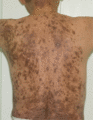
Figure 88.30
Post‐inflammatory hypermelanosis on the back following propranolol‐provoked lichenoid drug reaction.
Figure 88.34
Vitiligo. Epidermal sheet of marginal depigmented area showing marked reduction in the number of melanocytes.

Figure 88.38
Segmental vitiligo on the trunk seen under Wood's light. Note regressing congenital naevus in the centre of the affected area.
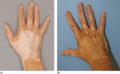
Figure 88.42
(a,b) Vitiligo: before and after camouflage of the hands.
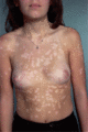
Figure 88.46
Hypopigmentation in a girl with resolving psoriasis.
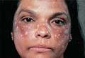
Figure 88.50
Depigmentation on the face following treatment of melasma with monobenzylether of hydroquinone. (Courtesy of St John's Dermatology Centre, London, UK...
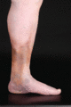
Figure 88.54
Haemosiderosis of the gaiter area in a 98‐year‐old man with longstanding venous insufficiency: note atrophie blanche above the medial malleolus.

Figure 88.58
Chrysiasis: mild lilac discoloration on the forehead and eyelids contrasting with the yellow of the elastotic skin on the bridge of the nose and eyebr...
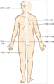
Figure 88.3
Regional variation in the distribution of epidermal melanocytes. The figures are mean values per mm 2 ± standard error of the mean. (From Rosdahl & R...

Figure 88.7
Diffuse hyperpigmentation with darkening of the hair and mucous membranes in a woman with Nelson syndrome following bilateral adrenalectomy.
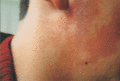
Figure 88.11
Erythromelanosis follicularis of the face and neck. (By permission of the copyright holder John Wiley and Sons, from Ermertcan et al . Erythromelanos...
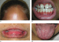
Figure 88.15
Addison disease: diffuse hypermelanosis of the skin (a) and gingivae (b) in a 13‐year‐old girl and of the labial mucosa (c) and tongue (d) of a 15‐yea...
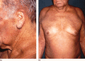
Figure 88.19
(a,b) Haemochromatosis: a 74‐year‐old male with gradual increase in skin pigmentation for 5 years. Extensive stippled skin pigmentation becoming confl...
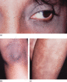
Figure 88.23
(a–c) Chloroquine pigmentation: patient received chloroquine for 9 years for rheumatoid arthritis and had developed increasing pigmentation of the per...
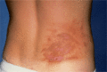
Figure 88.27
Phytophotodermatitis. Linear, streaky pigmentation following an acute blistering reaction caused by giant hogweed and sunlight.
Figure 88.31
Notalgia paraesthetica: note the circumscribed area of hypermelanosis near the base of the left scapula.
Figure 88.35
[Isomorphic or Koebner phenomenon at site of a scratch in a patient with vitiligo.
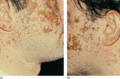
Figure 88.39
(a,b) Extensive vitiligo in a South Asian man: view of the sides of the face showing the convex expanding margins of the vitiliginous skin ‘eating int...
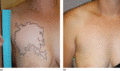
Figure 88.43
(a,b) Segmental vitiligo (a) before and (b) after treatment with non‐cultured epidermal cell transplantation.

Figure 88.47
Pityriasis alba.
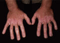
Figure 88.51
Occupational vitiligo due to tertiary‐butylphenol.
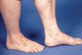
Figure 88.55
Haemosiderosis secondary to capillaritis which first erupted 6 months previously in a 57‐year‐old male.
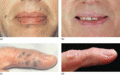
Figure 88.59
(a–d) Traumatic tattoo: accidental tattoo following explosion during school chemistry experiment; excellent response to Nd : YAG laser.
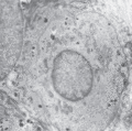
Figure 88.4
Melanocyte in the basal layer of the epidermis.

Figure 88.8
(a,b) Melasma in two female patients.
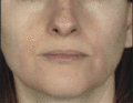
Figure 88.12
Peribuccal pigmentation of Brocq. (Courtesy and with permission of Dr Luciano Schiazza, Genoa, Italy.)
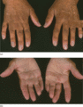
Figure 88.16
Nelson syndrome: hypermelanosis of the dorsa of the hands and of the palmar creases.
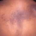
Figure 88.20
Macular amyloidosis: typical rippled hypermelanosis on the upper back of a 40‐year‐old Indian woman.

Figure 88.24
Chlorpromazine pigmentation: patchy bands of muddy pigmentation extending across the nose to the paranasal and preauricular skin in an elderly male re...
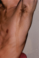
Figure 88.28
Berloque dermatitis.

Figure 88.32
Ashy dermatosis: views of two females aged 31 years (a,b) and 15 years (c) with multiple muddy grey non‐inflamed macules on the skin. (From Chang et ...

Figure 88.36
Typical distribution of vitiligo on the wrist and volar surface of the hand seen under Wood's light.
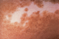
Figure 88.40
Trichrome vitiligo.
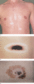
Figure 88.44
(a) Multiple halo naevi in a young man who also had vitiligo. (b) Unusually large halo naevus. (c) Halo phenomenon developing within a malignant melan...
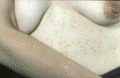
Figure 88.48
Hypomelanotic macules on a sun‐exposed arm compared with tan‐coloured macules on the trunk of the sun‐protected abdominal skin of a woman with pityria...
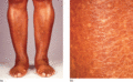
Figure 88.52
Typical appearances on the shins of a 57‐year‐old Afro‐Caribbean woman (a) with close‐up view illustrating discrete guttate hypomelanotic macules (b)....
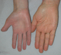
Figure 88.56
Carotenoderma: note yellowish hue of the palm on the right compared with the normal palm on the left. (Reproduced with permission and courtesy of the...

Figure 88.60
Lichenoid reaction in the red areas of a tattoo.
