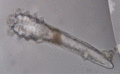
Figure 91.1
Demodex folliculorum mite showing its elongated worm‐like posterior body (opistostoma) and four sets of short legs on the upper body (podostoma). The...
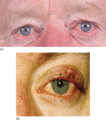
Figure 91.5
Ocular rosacea (OR). (a) Moderate (grade 2) OR with bilateral involvement, particularly of the lower eyelids. (b) More severe (grade 3) OR in this pat...
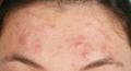
Figure 91.9
This patient with acne vulgaris has lesions that superficially resemble those of papulopustular rosacea (PPR). However, the lesions are larger and dee...
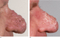
Figure 91.13
Rhinophyma before (a) and after (b) 4 months' treatment with isotretinoin.
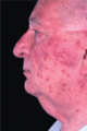
Figure 91.17
Corticosteroid‐induced rosacea‐like facial dermatosis.
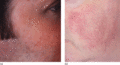
Figure 91.2
Erythematotelangiectatic rosacea (ETTR). (a) Facial erythema in moderate ETTR. (b) Prominent telangiectatic vessels on the lateral cheeks.
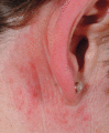
Figure 91.6
Grouped papules behind the ear of a patient with moderate papulopustular rosacea. This is a commonly overlooked location of inflammatory lesions.
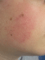
Figure 91.10
Pityriasis folliculorum in a young patient with localized area of erythema and fine scale.
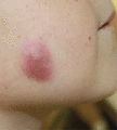
Figure 91.14
Idiopathic facial aseptic granuloma showing a well‐defined plum‐coloured nodule on the face of a 7‐year‐old boy. (From González Rodríguez et al . 201...
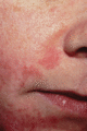
Figure 91.18
Perioral dermatitis.
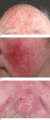
Figure 91.3
Papulopustular rosacea (PPR). (a) Papules and pustules on the forehead of a patient with PPR. (b) Erythema of the medial cheek area in a patient with ...
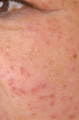
Figure 91.7
Granulomatous rosacea in a 55‐year‐old woman with a sudden onset of asymptomatic facial rash 4 months earlier. There is a profuse eruption of small, f...
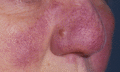
Figure 91.11
This patient with lupus pernio has skin changes that superficially resemble rosacea. Note the cyanotic hue more typical of sarcoid and the absence of ...
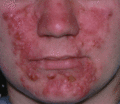
Figure 91.15
Rosacea fulminans showing an abrupt onset of severe inflammation with extensive pustule formation in a young woman.
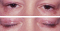
Figure 91.19
Periocular dermatitis.
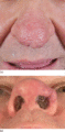
Figure 91.4
(a) Moderate to severe rhinophyma showing nasal distortion with a peau d'orange appearance of the prominent nasal follicles. (b) Swelling and distorti...
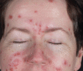
Figure 91.8
This woman was referred for management of ‘treatment‐resistant rosacea’. A skin biopsy showed non‐caseating granulomas. Investigations revealed her to...
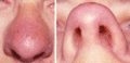
Figure 91.12
Solid facial lymphoedema involving predominantly the nose and mimicking rhinophyma. Note narrowing of the nares.
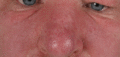
Figure 91.16
Solid facial lymphoedema is characterized by the presence of persistent, non‐tender, firm, upper facial swelling. Note the creases under the eyes in t...
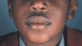
Figure 91.20
Childhood granulomatous periorificial dermatitis. (Courtesy of Professor Hywel Williams, University of Nottingham, UK.)
