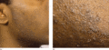
Figure 93.1
(a,b) Pseudofolliculitis barbae showing typical distribution (a) and close‐up view (b).
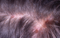
Figure 93.5
Scalp folliculitis with secondary excoriation.
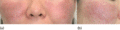
Figure 93.9
(a,b) Classical eosinophilic pustular folliculitis: well‐defined, dark erythematous plaques with numerous pustules and crusts involving the cheeks. (...
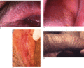
Figure 93.13
Fordyce spots on the vermilion of the upper lip (a), buccal mucosa (b), labia minora (c, courtesy of Dr Ekaterina Burova, Bedford Hospital, UK) and p...
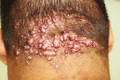
Figure 93.2
Folliculitis keloidalis of the nape of the neck. (Courtesy of Dr Ian Coulson, Burnley General Hospital, Burnley, Lancashire, UK.)
Figure 93.6
Follicular pustules on the anterior chest 48 h after irradiation with broadband UVA in a patient with actinic folliculitis.
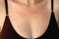
Figure 93.10
Immunodeficiency‐associated eosinophilic pustular folliculitis in 32‐year‐old woman with HIV infection: view of anterior chest. (Courtesy of Professo...
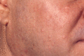
Figure 93.14
Sebaceous gland hyperplasia on the cheek of a 42‐year‐old man.
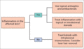
Figure 93.3
Treatment algorithm for folliculitis keloidalis.
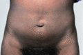
Figure 93.7
Disseminate and recurrent infundibulofolliculitis.
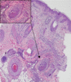
Figure 93.11
Infantile eosinophilic pustular folliculitis with dense infiltrate of eosinophils, spongiosis and microabscess formation within the follicle (inset). ...
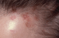
Figure 93.4
Varioliform scars at the scalp margin secondary to necrotizing lymphocytic folliculitis.
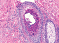
Figure 93.8
Histopathology of eosinophilic pustular folliculitis showing dense accumulation of eosinophils within the follicular canal. (Courtesy of Professor Lu...

Figure 93.12
Sterile pustules in the scalp of a 23‐month‐old boy with infantile eosinophilic pustular folliculitis. (Reproduced from Alonso‐Castro et al . 2012 [...
