Figure 95.1
Longitudinal section of a digit showing the dorsal nail apparatus.

Figure 95.5
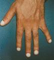
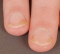
Clubbing: Lovibond's profile sign. The angle is normally less than 160° but exceeds 180° in clubbing.

Figure 95.9
Pincer nail.

Figure 95.13
Nail pterygium due to lichen planus.

Figure 95.17
Trachyonychia: roughened surface of up to 20 nails.

Figure 95.21
Green pigmentation of onycholytic fingernail due to Pseudomonas .

Figure 95.25
(a) Longitudinal erythronychia. (b) The longitudinal ridge in the nail bed corresponds to the groove on the undersurface of the nail plate.

Figure 95.29
Nail biting can be extensive, with damage to the nail folds and nail plate causing subungual haemorrhage.

Figure 95.33
(a,b) Ingrowing great toenail complicated by proximal ingrowing (retronychia).

Figure 95.37
This lesion was mistaken for an ingrowing toenail. X‐rays confirmed the presence of subungual exostosis.

Figure 95.41
Submatricial fibrokeratoma pressing onto the underlying matrix with subsequent longitudinal smooth groove.

Figure 95.45
X‐rays showing massive osteolysis of the distal bony phalanx associated with subungual keratoacanthoma.

Figure 95.49
Superficial fibromyxoma of the nail bed elevating the distal plate.

Figure 95.53
Narrow longitudinal melanonychia on a thumb. Dermoscopy showed loss of parallelism that prompted excisional biopsy. Histological examination revealed ...

Figure 95.57
(a–c) Chronic paronychia: paronychial swelling, loss of cuticle and mildly dystrophic nail in early disease (a,b); severe nail dystrophy in more advan...

Figure 95.61
Psoriasis: pitting.

Figure 95.65
Multiple transverse grooves of the thumbnails.

Figure 95.69
Lichen planus with longitudinal melanonychia.

Figure 95.73
Ultrasound: grey scale ultrasound (longitudinal view) demonstrates the normal sonographic anatomy of the nail (a); 3D ultrasound reconstruction of the...

Figure 95.77
Normal nail fold capillaries (×60) (a); acrocyanosis showing dilatation of the nail fold capillaries, stasis and thrombosis of many vessels (b); rheum...

Figure 95.81
(a) Avulsion of the proximal third of the plate demonstrates that the pigment area responsible for the pigmentation extends longitudinally on the matr...

Figure 95.85
(a) Dermatophyte onychomycosis with longitudinal spikes. (b) After surgical removal of the yellow streaks.

Figure 95.89
(a) Ingrowing toenail with pyogenic granuloma. (b) After curettage of the pyogenic granuloma, a lateral strip of nail is avulsed. (c) A cotton‐tipped ...

Figure 95.2
Direction of differentiation and cell movement within the nail apparatus.

Figure 95.6
Clubbing: Curth's modified profile sign.

Figure 95.10
Anonychia: nail loss in a 50‐year‐old man with dominant dystrophic epidermolysis bullosa.

Figure 95.14
Ventral pterygium due to allergy to formaldehyde nail hardener.

Figure 95.18
Onychoschizia (lamellar splitting).

Figure 95.22
Total leukonychia.

Figure 95.26
Transverse ridges resulting from habit tic.

Figure 95.30
Early onychogryphosis of the left great toenail.

Figure 95.34
Pyogenic granuloma resulting from friction of the overlapping second toe against the lateral aspect of the great toenail.

Figure 95.38
Digital myxoid pseudocyst type A.

Figure 95.42
Intraungual (dissecting) fibrokeratoma. The lesion grows within the nail plate and emerges at its distal half.

Figure 95.46
Onychomatricoma, pigmented variant: note the very well‐delimited longitudinal thickening of the plate.

Figure 95.50
Onychopapilloma: note the longitudinal erythronychia starting in the distal matrix, the distal splinter haemorrhages and the onycholysis at its distal...

Figure 95.54
Friable granulation tissue under the plate of the great toenail in an old lady wearing sandals all year round. Pyogenic granuloma was suspected but hi...

Figure 95.58
Painful dorsolateral fissure of the fingertip.

Figure 95.62
Psoriasis: salmon patches progressing to onycholysis.

Figure 95.66
Acropustulosis: nail plate has been destroyed by intense pustular inflammation.

Figure 95.70
Anonychia following lichen planus.

Figure 95.74
Nail melanoma: clinical presentation (a); dermoscopy (b) and in vivo reflectance confocal microscopy (c) of the nail matrix in the same patient. (C...

Figure 95.78
Dermatomyositis showing dilated nail folds capillaries and obstructed and thrombosed capillaries (×60) (inset).

Figure 95.82
(a) Avulsion of the proximal third of the plate exposes the wide pigment area responsible for the longitudinal pigmentation. (b) An incision is carrie...

Figure 95.86
(a) Chronic paronychia resistant to topicals and steroid injections. (b) Crescent‐shaped excision of a part of the proximal nail fold. (c) Complete re...

Figure 95.90
Allergy to nail varnish presenting as an eyelid dermatitis.

Figure 95.3
Arterial supply of the distal finger.

Figure 95.7
Schamroth's window is seen clearly in this image of normal nails.

Figure 95.11
Onycholysis: idiopathic type.

Figure 95.15
Median canaliform dystrophy of Heller.

Figure 95.19
Longitudinal ridging of the nail.

Figure 95.23
Punctate leukonychia.

Figure 95.27
Median canaliform dystrophy of Heller affecting distal portion of nail plate; the enlarged lunula and transverse ridging seen proximally reflect chron...

Figure 95.31
Severe onychogryphosis resulting from neglect.

Figure 95.35
Painful glomus tumour of the nail bed. Note the bluish hue.

Figure 95.39
Digital myxoid pseudocyst type B. Note the longitudinal groove arising from underneath the proximal nail fold where the matrix is compressed by the ov...

Figure 95.43
Multiple soft fibrokeratomas in tuberous sclerosis.

Figure 95.47
Onychomatricoma: ‘woodworm’ cavities in the nail plate are especially visible in this longstanding case (>40 years).

Figure 95.51
Bowen disease: warty lesion of the distal bed and hyponychium. The lesion was treated for several years as a wart.

Figure 95.55
Acute bacterial paronychia (whitlow).

Figure 95.59
Hangnail.

Figure 95.63
Psoriasis: distal onycholysis.

Figure 95.67
Darier disease: white and red longitudinal lines and distal notching.

Figure 95.71
Paronychia of the little finger in a 2‐year‐old child.

Figure 95.75
Axial T2‐weighted image at the level of the distal interphalangeal joint (arrows): pedicle of the myxoid pseudocyst connected with the joint (arrows) ...

Figure 95.79
Lupus erythematosus (a) and systemic sclerosis (b). (Courtesy of the copyright holder C. Mathis, Belgium.)

Figure 95.83
Lateral longitudinal biopsy. Note the sigmoid shape of the defect that can be easily closed.

Figure 95.87
Two lateral incisions at 45° allow reflection of the proximal nail fold; visualization of the tumour and its removal.

Figure 95.91
Staining of the nail plates from nail varnish.

Figure 95.4
Proliferating epithelial cells of the matrix and ventral aspect of the proximal nail fold, staining with the antibody MIB‐1.

Figure 95.8
Koilonychia. This image is of congenital koilonychia in a young girl.

Figure 95.12
Photo‐onycholysis with a uniform pattern of discoloured onycholysis in the midline.

Figure 95.16
Beau's lines present as transverse grooves in the nail matching the proximal margin of the nail matrix and lunula.

Figure 95.20
Orange pigmentation of onycholytic toenail due to orange dye from work‐boots.

Figure 95.24
Yellow nail syndrome

Figure 95.28
Chronic paronychia.

Figure 95.32
(a–c) Onychogryphosis is often best treated with ablation of the nail matrix.

Figure 95.36
Subungual exostosis: exophytic growth of bone emerging from under the nail plate through collarette of skin (note the telangiectases) (a); exostosis ...

Figure 95.40
Digital myxoid pseudocyst type C. Note the red macule within the lunula.

Figure 95.44
Subungual distal keratoacanthoma. Note the keratotic plug on the distal bed.

Figure 95.48
Onychomatricoma: showing the sea anemone‐like matrix tumour and the cavities in the avulsed nail plate into which digitate projections from the tumour...

Figure 95.52
Onycholysis and oozing of the great toenail bed due to invasive squamous cell carcinoma.

Figure 95.56
Herpetic whitlow.

Figure 95.60
Subungual abscess in neutropenic patient receiving cancer chemotherapy. (Courtesy of B. Fouilloux, France.)

Figure 95.64
Psoriasis: subungual hyperkeratosis.

Figure 95.68
Severe onychatrophy from juvenile onset lichen planus of nails.

Figure 95.72
Transverse acro‐osteolysis of the fingernail (a); acro‐osteolysis of the toenail (b). (Courtesy of J. L. Drapé, France.)

Figure 95.76
Sagittal section T2 fat saturated image: pedicle connecting with the joint (arrow) under the extensor tendon (arrow heads). (Courtesy of the copyrigh...

Figure 95.80
(a) Avulsion of the proximal third of the plate exposes the pigment area responsible for the longitudinal pigmentation. (b) A 3 mm punch is performed ...

Figure 95.84
Lateral avulsion (‘sardine tin’ avulsion) allows exposure of the complete nail bed and excisional biopsy of the nail bed tumour.

Figure 95.88
Trap door avulsion permits access to the nail bed tumour. The latter will be removed in a longitudinal excision.

Figure 95.92
Complication of nail extensions: allergy to acrylate adhesive presenting as onycholysis.

