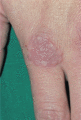
Figure 97.1
Granuloma annulare on the dorsum of the hand – a typical site.
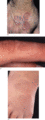
Figure 97.5
Common sites of localized granuloma annulare. (a) On the dorsum of the hand; note the atrophy in the centre of the lesions. (b) On the dorsum of a fin...
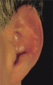
Figure 97.9
Granuloma annulare on the ear. Note the nodules overlying the auricular cartilage of the antihelix.
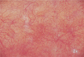
Figure 97.13
Prominent telangiectasia in an area of necrobiosis.
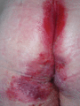
Figure 97.17
Severe perianal Crohn disease.
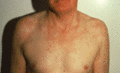
Figure 97.2
Generalized granuloma annulare showing a clear photo‐distribution over the ‘V’ of the neck and shoulders.
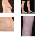
Figure 97.6
Common sites of generalized granuloma annulare. (a) Right flank. (b) Abdomen. (c, d) Involvement of the lower extremities.
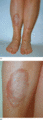
Figure 97.10
(a) Necrobiosis lipoidica of the shins in a 50‐year‐old woman. (b) Close‐up view of the shin.
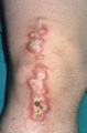
Figure 97.14
Necrobiosis lipoidica Koebnerizing in a scar from previous knee surgery.
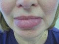
Figure 97.18
Lip swelling due to oro‐facial granulomatosis developed in this patient some 5 years before she presented with severe intestinal Crohn disease. (Cour...
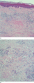
Figure 97.3
(a) Necrobiotic palisading granuloma showing well‐circumscribed central necrobiosis surrounded by a palisade of histiocytes. (b) Close‐up view showing...
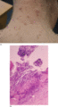
Figure 97.7
(a) Perforating granuloma annulare on the neck and (b) histology showing transepidermal elimination of necrobiotic collagen.
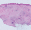
Figure 97.11
Necrobiosis lipoidica showing extensive necrobiosis in the dermis.
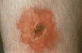
Figure 97.15
Ulcerated necrobiosis lipoidica in a 34‐year‐old woman with diabetes.
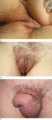
Figure 97.19
(a) Vulval swelling due to Crohn disease presented in the same patient as shown in Figure about 1 year after she developed intestinal Crohn disease....
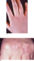
Figure 97.4
(a) Typical appearance of localized granuloma annulare over the knuckles. (b) Appearance of granuloma annulare over the knuckles on clenching the fist...
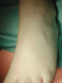
Figure 97.8
Subcutaneous granuloma annulare with nodules visible on the dorsolateral aspect of a child's foot.
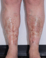
Figure 97.12
Chronic necrobiosis lipoidica (over a 20‐year period) in a 41‐year‐old patient with insulin‐dependent diabetes. Note the marked atrophy and telangiect...
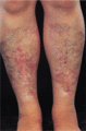
Figure 97.16
‘Burnt out’ necrobiosis lipoidica – marked atrophy is evident.
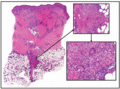
Figure 97.20
Cutaneous Crohn disease showing superficial and deep perivascular dermal infiltrates and a predominantly septal panniculitis. At higher magnification ...
