Figure 144.1
Dermoscopic naevi patterns.
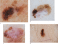
Figure 144.5
(a) Dermoscopic image of a melanoma on the lower back revealing an atypical network (solid box), regression structures including granularity and scar‐...
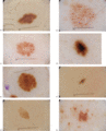
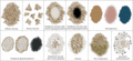
Figure 144.2
(a) Reticular diffuse naevus. (b) Reticular patchy naevus. (c) Peripheral reticular with central hypopigmentation naevus. (d) Peripheral reticular wit...
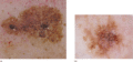
Figure 144.6
(a) Dermoscopic image of a lentigo maligna located on the nose with perifollciular granularity and asymmetrical grey perifollicular openings (solid bo...
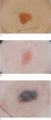
Figure 144.3
Homogeneous pattern naevi: (a) homogeneous brown; (b) homogeneous pink; and (c) homogeneous grey‐blue.
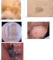
Figure 144.7
(a, b) Dermoscpic images of melanomas located on the soles with a parallel ridge pattern. (c) Dermoscopic image (c1) of a melanoma, 0.6 mm in thicknes...
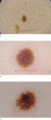
Figure 144.4
Naevi with peripheral globules: (a) growing naevus with a single row of globules; (b) Spitz naevus with multiple rows of globules; and (c) Spitz naevu...
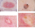
Figure 144.8
(a) Dermoscopic image of an amelanotic melanoma that is not of the nodular subtype, showing serpentine vessels, dotted vessels and vascular blush thro...
