Figure 31.1
Structure of HIV. (Adapted from AIDS Info 2014 [ ].)

Figure 31.5
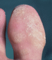
Severe plantar psoriasis in HIV infection. (Courtesy of Medical Illustration UK Ltd, Chelsea and Westminster Hospital, London, UK.)

Figure 31.9
Bacillary angiomatosis: purple nodules on the face. (Courtesy of Medical Illustration UK Ltd, Chelsea and Westminster Hospital, London, UK.)

Figure 31.13
Cytomegalovirus vasculitis: leg ulcers. (Courtesy of Medical Illustration UK Ltd, Chelsea and Westminster Hospital, London, UK.)

Figure 31.17
Atypical mollusca: flesh‐coloured papules and nodules on the forehead. (Courtesy of Media Resources UCL, London, UK.)

Figure 31.21
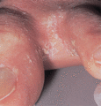
Norwegian scabies: interdigital scale.

Figure 31.25
Alopecia areata. (Courtesy of Medical Illustration UK Ltd, Chelsea and Westminster Hospital, London, UK.)

Figure 31.29
Cytomegalovirus immune restoration disease: necrotizing impetiginized ulcer on the left ear. (Courtesy of Imperial College School of Medicine, London...

Figure 31.2
Stages in the HIV life‐cycle (blue ovals) showing location, interaction with, and selective pressure of candidate cellular host factors/proteins that ...

Figure 31.6
Eosinophilic folliculitis: excoriated papules on the trunk. (Courtesy of Medical Illustration UK Ltd, Chelsea and Westminster Hospital, London, UK.)

Figure 31.10
Bacillary angiomatosis: Warthin–Starry silver staining of Bartonella bacilliformis and proliferative vascular channels. (Courtesy of Dr N. Francis,...

Figure 31.14
Cytomegalovirus infection: nodular prurigo‐like eruption on the back. (Courtesy of Medical Illustration UK Ltd, Chelsea and Westminster Hospital, Lon...

Figure 31.18
Tinea corporis and faciei. (Courtesy of Medical Illustration UK Ltd, Chelsea and Westminster Hospital, London, UK.)

Figure 31.22
Kaposi sarcoma. (a) Purple nodules on the palate. (b) Multiple purple nodules and plaques on the back. (Courtesy of Medical Illustration UK Ltd, Chel...

Figure 31.26
Hairy leukoplakia. (Courtesy of Media Resources UCL Trust, London, UK.)

Figure 31.3
Thrombocytopenic purpura: purpuric macule on the finger. (Courtesy of Medical Illustration UK Ltd, Chelsea and Westminster Hospital, London, UK.)

Figure 31.7
Granuloma annulare on the hand. (Courtesy of Media Resources UCL, London, UK.)

Figure 31.11
Chronic perianal ulceration in herpes simplex infection before the era of highly active antiretroviral therapy.

Figure 31.15
Human papillomavirus infection: myrmecia on the great toe. (Courtesy of Media Resources UCL, London, UK.)

Figure 31.19
Cryptococcosis. (a) Necrotizing papules and nodules on the right ear and neck. (b) Close up of necrotizing papules. (Courtesy of Medical Illustration...

Figure 31.23
Squamous carcinoma. (a) Ulcerated nodule on the right upper eyelid. (b) Metastatic zosteriform ulceration of the left axilla and chest. (Courtesy of ...

Figure 31.27
Cheilitis caused by indinavir (IDV, INR). (Courtesy of Imperial College School of Medicine, London, UK.)

Figure 31.4
Seborrhoeic dermatitis on the face. (Courtesy of Medical Illustration UK Ltd, Chelsea and Westminster Hospital, London, UK.)

Figure 31.8
Staphylococcal scalded skin syndrome: staphylococcal pneumonia in an HIV‐positive intravenous drug addict. (Courtesy of Medical Illustration UK Ltd, ...

Figure 31.12
Chronic verrucous herpes zoster. (Courtesy of Medical Illustration UK Ltd, Chelsea and Westminster Hospital, London, UK.)

Figure 31.16
Epidermodysplasia verruciformis in human papillomavirus infection: discrete and confluent warty papules on the right cheek and neck. (Courtesy of Imp...

Figure 31.20
Penicilliosis (Courtesy of Professor Vesarat Wessagowit, Bangkok, Thailand.)

Figure 31.24
Melanoma: 2‐month history of rapidly growing amelanotic nodule on the left arm; Breslow thickness >10 mm. (Courtesy of Medical Illustration UK Ltd, C...

Figure 31.28
Herpes simplex immune restoration disease: chronic erosions on the penis. (Courtesy of Medical Illustration UK Ltd, Chelsea and Westminster Hospital,...

