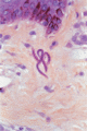
Figure 33.1
Microfilaria of Onchocerca volvulus in the upper dermis. Note the absence of inflammation around the live organism. (Courtesy of Dr C. McDougall, S...
Figure 33.5
Late lichenified onchodermatitis (pachyderma).
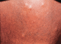
Figure 33.9
Streptocerciasis. Pigmentary changes and nodules in a patient from Zaire. (Courtesy of the Armed Forces Institute of Pathology, Washington, DC, USA.)...
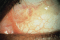
Figure 33.13
Loa loa . Creamy white adult worm crossing the conjunctiva of a Nigerian patient.
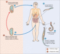
Figure 33.17
Life cycle of Enterobius vermicularis . (Courtesy of the Centers for Disease Control and Prevention. http://www.cdc.gov/dpdx/dracunculiasis/index.h...
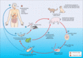
Figure 33.21
Life cycle of Gnathostoma spinigerum and G. hispidum . (Courtesy of the Centers for Disease Control and Prevention http://www.cdc.gov/parasites/g...
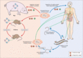
Figure 33.25
Life cycle of Trichinella . (Courtesy of the Centers for Disease Control and Prevention http://www.cdc.gov/parasites/trichinellosis/biology.html l...
Figure 33.29
Schistosoma haematobium ovum in urine. (Courtesy of Mr A.H. Moody, Hospital for Tropical Diseases, London, UK.)
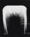
Figure 33.33
Calcified cysticercal cysts in the muscles of the thighs and pelvis.
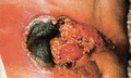
Figure 33.37
Cutaneous amoebiasis. Rapidly spreading ulcer around a colostomy in a patient with intestinal amoebiasis. (Courtesy of Meddia (Medical and Diagnostic...
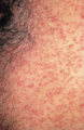
Figure 33.41
Trypanosomal rash (same patient as Figure ). The maculopapular rash is becoming haemorrhagic.
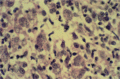
Figure 33.45
Cutaneous leishmaniasis (high power, H&E); biopsy of early lesion, showing abundant amastigotes in macrophages, and a few plasma cells. (Courtesy of ...
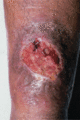
Figure 33.49
Cutaneous leishmaniasis due to Leishmania major from Sudan. An ulcer with a raised edge.
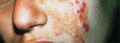
Figure 33.53
Leishmaniasis recidivans (lupoid leishmaniasis) from Baghdad, showing active papules cropping in the edge of the scar of the healed sore. (Courtesy o...
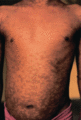
Figure 33.57
Post‐kala‐azar dermal leishmaniasis in an Indian person showing the extensive hypopigmented macular rash.
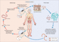
Figure 33.2
Life cycle of Onchocera volvulus . (Courtesy of the Centers for Disease Control and Prevention. http://www.cdc.gov/dpdx/onchocerciasis/ last acces...
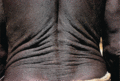
Figure 33.6
Late onchocerciasis. Atrophy of skin and damage to supporting tissue cause the skin to sag in folds (lizard skin).
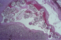
Figure 33.10
Lymphatic filariasis (low power, H&E). Female adult Wuchereria bancrofti within lymph node sinus. Multiple cross‐sections of the worm are seen. (Co...
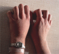
Figure 33.14
Calabar swelling of the dorsum of the left hand in a European patient with loaiasis. In this case no microfilariae were seen in the blood.
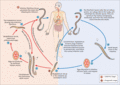
Figure 33.18
The life cycle of Strongyloides stercoralis . (Courtesy of the Centers for Disease Control and Prevention http://www.cdc.gov/parasites/strongyloide...
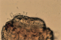
Figure 33.22
Gnathostoma spinigerum . Head of an adult worm that was extruded from a subcutaneous swelling in a patient from Bangladesh. The crown of hooklets is c...
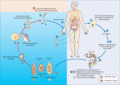
Figure 33.26
Life cycle of the Schistosoma . (Courtesy of the Centers for Disease Control and Prevention http://www.cdc.gov/parasites/schistosomiasis/biology.ht...
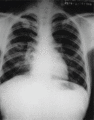
Figure 33.30
Paragonimiasis. Chest X‐ray showing three discrete opacities, in two of which cavitation is present, characteristic of the disease in Thailand. (Cour...
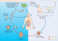
Figure 33.34
Life cycle of spargana. (Courtesy of the Centers for Disease Control and Prevention http://www.cdc.gov/dpdx/sparganosis/ last accessed November 201...

Figure 33.38
African trypanosomiasis parasite life cycle. (Courtesy of the Centers for Disease Control and Prevention http://www.cdc.gov/parasites/sleepingsickne...

Figure 33.42
Chagas disease. Unilateral oedema of the eyelids and orbit (Chagas–Mazza– Romaña's sign). (Courtesy of Professor M. Miles, London School of Hygiene a...
Figure 33.46
Chronic cutaneous leishmaniasis. Granulomatous dermatitis without necrosis. There are no amastigotes to be found in this case, and the differential di...
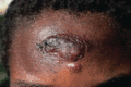
Figure 33.50
Cutaneous leishmaniasis due to Leishmania aethiopica from Kenya. A large nodule with many satellite papules and abundant parasites.
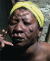
Figure 33.54
Diffuse cutaneous leishmaniasis due to Leishmania aethiopica in Ethiopia. The face is covered with infiltration and nodulation but there is no ulcer...
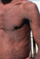
Figure 33.58
Post‐kala‐azar dermal leishmaniasis in an Indian person. A later stage than that shown in Figure . Papules are beginning to develop.
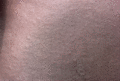
Figure 33.3
Acute papular onchodermatitis in a Nigerian. Early in the disease the papules are usually urticarial.
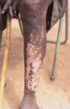
Figure 33.7
Depigmentation over the shin in late onchocerciasis (leopard skin). This is common in African endemic areas. Note the stick for the blind.
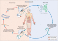
Figure 33.11
Life cycle of Wuchereria bancrofti . (Courtesy of the Centers for Disease Control and Prevention. http://www.cdc.gov/dpdx/onchocerciasis/ last acc...
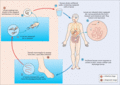
Figure 33.15
Life cycle of Dracunculus medinensis . (Courtesy of the Centers for Disease Control and Prevention. http://www.cdc.gov/dpdx/dracunculiasis/index.ht...
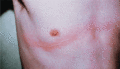
Figure 33.19
Larva currens rash of Strongyloides stercoralis . The weal and flare will have disappeared in a few hours.
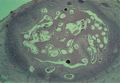
Figure 33.23
Dirofilariasis. Low‐power view of cold abscess from the cheek. It shows degenerate coiled Dirofilaria worm within an abscess, and surrounding lympho...
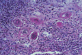
Figure 33.27
Schistosomiasis of the vulva (medium power, H&E) showing six schistosome eggs with surrounding lymphoplasmocytic infiltration. (Courtesy of Professor...
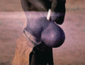
Figure 33.31
Hydatid disease. Subcutaneous hydatid cyst presenting as a hernia, in a woman from Turkana, Kenya. (Courtesy of Dr C.N.L. MacPherson, Liverpool Schoo...
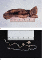
Figure 33.35
(a) Fibrous encapsulated cold abscess from the groin, which contained (b) a 22‐cm long sparganum. (Courtesy of Professor S.B. Lucas, King's College L...
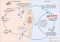
Figure 33.39
American trypanosomiasis parasite life cycle. (Courtesy of the Centers for Disease Control and Prevention http://www.cdc.gov/parasites/chagas/biolog...
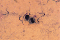
Figure 33.43
Trypanosomes in a thick blood film, stained with Giemsa. (Courtesy of Dr P.L. Chiodini, Hospital for Tropical Diseases, London, UK.)
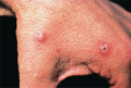
Figure 33.47
Cutaneous leishmaniasis due to Leishmania major : early papules, one of which is starting to show central crusting.
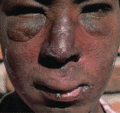
Figure 33.51
Nasal involvement, and marked inflammatory oedema in leishmaniasis due to Leishmania aethiopica in Ethiopia.
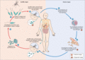
Figure 33.55
Life cycle of leishmaniasis parasite. (Courtesy of the Centers for Disease Control and Prevention http://www.cdc.gov/parasites/leishmaniasis/biology...
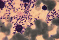
Figure 33.59
Amastigotes of Leishmania infantum in a bone marrow smear from a patient with visceral leishmaniasis and AIDS. (Courtesy of Mr A.H. Moody, Hospital...
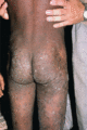
Figure 33.4
Chronic papular onchodermatitis. Lesions are starting to lichenify.
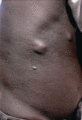
Figure 33.8
Cutaneous oedema and cutaneous and subcutaneous nodules in a Nigerian with onchocerciasis.
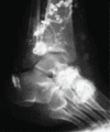
Figure 33.12
Lymphangiogram showing tortuous dilated lymphatic vessels in the leg of a patient with lymphatic filariasis from Mauritius.
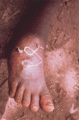
Figure 33.16
Guinea worm. Adult female Dracunculus medinensis emerging from the skin of a Nigerian patient. (Courtesy of Dr R. Muller, CAB International Institu...
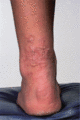
Figure 33.20
Cutaneous larva migrans (creeping eruption). There are several tortuous indurated inflamed worm tracks, in some of which may be seen a blister that ma...
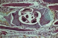
Figure 33.24
Trichinosis. Medium‐power view, Movat stain, showing Trichinella worm within a ‘nurse cell’ in skeletal muscle. There is surrounding oedema, muscle ...
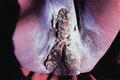
Figure 33.28
Schistosomiasis of the vulva and anus. Condylomatous lesions containing granulomas around schistosome ova. (Courtesy of Professor G. Nelson, Liverpoo...
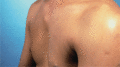
Figure 33.32
Cysticercosis in a man from North India. A subcutaneous cyst is seen over the sternum. (Courtesy of Dr A.P. Hall, Hospital for Tropical Diseases, Lon...
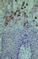
Figure 33.36
Cutaneous amoebiasis. High‐power view of skin biopsy, showing Entamoeba histolytica trophozoites on the epidermis, causing necrosis. The amoebae are...
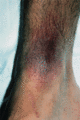
Figure 33.40
Trypanosomal chancre due to Trypanosoma rhodesiense infection, appearing 6 days after the bite of the tsetse fly. The lesion is swollen, inflamed an...
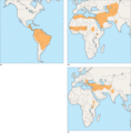
Figure 33.44
(a) Geographical distribution of cutaneous and mucocutaneous leishmaniasis in the New World. (b) Geographical distribution of Old World cutaneous leis...
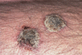
Figure 33.48
Cutaneous leishmaniasis due to Leishmania major from Saudi Arabia, showing marked and persistent crusting.
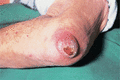
Figure 33.52
Cutaneous leishmaniasis due to Leishmania infantum , from Spain. The fleshy nodule, with relatively little inflammation, is characteristic.
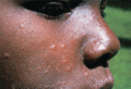
Figure 33.56
Post‐kala‐azar dermal leishmaniasis. Typical facial papules in a Kenyan arising 6 weeks after treatment and healing spontaneously. (Courtesy of Dr J....
