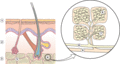
Figure 99.1
Schematic representation of the anatomy of subcutaneous fat with detailed view showing the vascular supply to the fat lobule and its constituent micro...
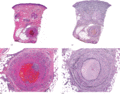
Figure 99.5
Histopathological features of superficial thrombophlebitis. (a) Scanning view showing involvement of a large vein in the septa of subcutaneous tissue....
Figure 99.9
Histopathological features of necrobiosis lipoidica extending to subcutaneous tissue. (a) Scanning power showing involvement of the full thickness of ...
Figure 99.13
Histopathological findings in subcutaneous granuloma annulare. (a) Scanning power showing the involvement of deeper dermis and subcutaneous tissue. (b...
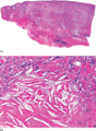
Figure 99.17
Histopathological features of necrobiotic xanthogranuloma.
(a) Scanning power showing diffuse involvement of the entire thickness of the dermis and ex...
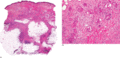
Figure 99.21
Histopathological features of a late stage lesion of erythema nodosum. (a) Scanning power showing a mostly septal panniculitis. (b) Numerous multinucl...
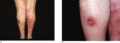
Figure 99.25
Clinical features of erythema induratum. (a) Erythematous nodules and plaques on the posterior aspect of the legs of an adult woman. (b) Some of the l...
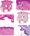
Figure 99.29
Histopathological findings in sclerosing panniculitis. (a) Scanning power showing thickened septa and cystic spaces replacing the fat lobules. (b) A c...
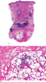
Figure 99.33
Histopathology of equestrian panniculitis. (a) Scanning power showing dense nodular infiltrates in deeper dermis. (b) Small aggregate of lymphocytes i...
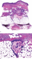
Figure 99.37
Histopathological features of pancreatic panniculitis. (a) Scanning power showing a mostly lobular panniculitis. (b) A group of ‘ghost’ adipocytes sur...
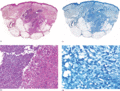
Figure 99.41
Histopathological features in a cutaneous infection by M. chelonae . (a) Scanning power showing involvement of the reticular dermis and subcutaneous ...

Figure 99.45
Histopathological features of neutrophilic lobular panniculitis. (a) Scanning power showing a predominantly lobular panniculitis. (b) The lobular infi...

Figure 99.49
Histopathological features of encapsulated fat necrosis. (a) Scanning power showing an enucleated and very well‐circumscribed lesion. (b) Necrotic adi...
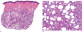
Figure 99.53
Histopathological features of subcutaneous fat necrosis of the newborn. (a) Scanning power showing a lobular panniculitis. (b) The fat lobule is repla...
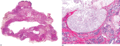
Figure 99.57
Histopathology of gouty panniculitis. (a) Scanning power showing involvement of the subcutaneous tissue, whereas the dermis is spared. (b) Basophilic ...
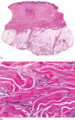
Figure 99.61
Histopathological features of sclerosing postirradiation panniculitis. (a) Scanning power showing a mostly lobular panniculitis. (b) Sclerotic collage...
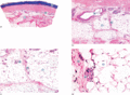
Figure 99.2
(a) Scanning power view of the normal skin of the sole. The epidermis is covered by a thick compact orthokeratotic horny layer (star). Numerous eccrin...
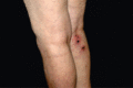
Figure 99.6
Clinical appearance of cutaneous polyarteritis nodosa showing livedo reticularis of the lower extremities with ulcerated nodules on the right calf of ...
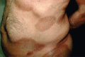
Figure 99.10
Morphoea profunda. The lesions consisted of indurated, hyperpigmented and slightly depressed plaques.
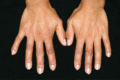
Figure 99.14
Rheumatoid nodules involving the dorsum of the fingers in an adult woman with seropositive rheumatoid arthritis.
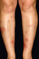
Figure 99.18
Characteristic eruption of erythema nodosum with lesions in different stages of evolution involving the anterior aspect of the legs of an adult woman....
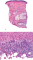
Figure 99.22
Histopathological findings of a very late stage lesion of erythema nodosum. (a) Scanning power showing very thick connective tissue septa at the subcu...
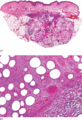
Figure 99.26
Histopathological features of an early lesion of erythema induratum. (a) Scanning power showing a mostly lobular panniculitis. (b) Higher magnificatio...
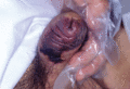
Figure 99.30
Calciphylaxis involving the penis in a patient with end‐stage renal disease.
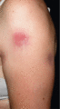
Figure 99.34
Clinical features of lupus panniculitis showing an active erythematous subcutaneous nodule and areas of hyperpigmented lipoatrophy secondary to regres...
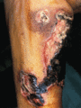
Figure 99.38
Panniculitis associated with α 1 ‐antitrypsin deficiency. Necrotic ulcers exudate oily material that result from necrotic adipocytes.
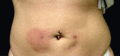
Figure 99.42
Panniculitis on the anterior abdominal wall secondary to subcutaneous glatiramer acetate injections for the treatment of multiple sclerosis.
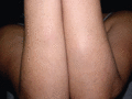
Figure 99.46
Subcutaneous sarcoidosis. Subcutaneous firm nodules on the forearms covered by normal appearing skin.
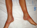
Figure 99.50
Lipoatrophic panniculitis of the ankles in a 12‐year‐old boy.
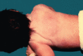
Figure 99.54
Sclerema neonatorum in a newborn with generalized woody induration of the skin.
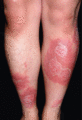
Figure 99.58
Subcutaneous pannicultis‐like T‐cell lymphoma. Erythematous plaques involving the anterior aspects of the lower extremities.
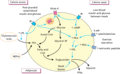
Figure 99.3
Simplified outline of lipogenesis in an adipocyte during energy excess and lipolysis during calorie need. Effects of hormones and enzymes are in blue....
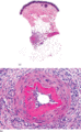
Figure 99.7
Histopathological features of cutaneous polyarteritis nodosa. (a) Scanning view of a punch biopsy showing involvement of a vessel of the septa of subc...
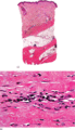
Figure 99.11
Histopathology of deep morphoea. (a) Scanning power showing sclerosis of the deeper reticular dermis and the septa of subcutaneous tissue. Note that t...
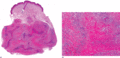
Figure 99.15
Histopathological features of rheumatoid nodule. (a) Scanning power showing a diffuse replacement of subcutaneous tissue by a fibrotic process with sc...
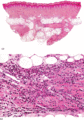
Figure 99.19
Histopathological features of an early lesion of erythema nodosum. (a) Scanning power showing a mostly septal panniculitis with thickened connective t...
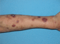
Figure 99.23
Clinical features of erythema nodosum leprosum. Acute onset of erythematous nodules involving the left upper extremity in a patient with lepromatous l...
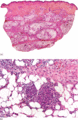
Figure 99.27
Histopathological features of a fully developed lesion of erythema induratum. (a) Scanning power showing a mostly lobular panniculitis. (b) Small gran...
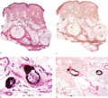
Figure 99.31
Histopathological features of calciphylaxis. (a) Scanning power view showing involvement of the vessels of deeper reticular dermis. (b) Calcification ...
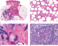
Figure 99.35
Histopathological features of lupus panniculitis. (a) Scanning magnification showing a predominantly lobular panniculitis. (b) Dense lymphoid aggregat...
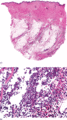
Figure 99.39
Histopathological features of α 1 ‐antitrypsin deficiency panniculitis. (a) Scanning power showing involvement at the septa and the periphery of the f...
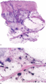
Figure 99.43
Histopathological features of panniculitis secondary to subcutaneous glatiramer acetate injections for the treatment of multiple sclerosis. (a) Scanni...
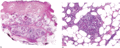
Figure 99.47
Histopathological features of subcutaneous sarcoidosis. (a) Scanning power showing a predominantly lobular panniculitis. (b) Small non‐caseating granu...
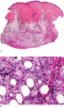
Figure 99.51
Histopathological findings in lipoatrophic panniculitis of the ankles in children. (a) Scanning power showing a mostly lobular panniculitis. (b) The f...
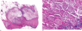
Figure 99.55
Histopathological features of sclerema neonatorum. (a) Scanning power showing necrosis of the entire fat lobules and thickened connective tissue septa...
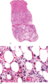
Figure 99.59
Histopathological features of subcutaneous panniculitis‐like T‐cell lymphoma. (a) Scanning power showing features simulating a lobular panniculitis. (...
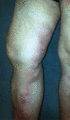
Figure 99.4
Superficial thrombophlebitis. Varicosities and erythematous nodules with linear arrangement involving the right lower extremity.
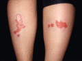
Figure 99.8
Necrobiosis lipoidica showing yellowish indurated plaques on the anterior aspect of the legs in a diabetic woman.
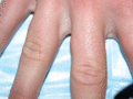
Figure 99.12
Subcutaneous granuloma annulare involving the lateral aspect of the first phalanx of the third right finger in a 14‐year‐old boy.
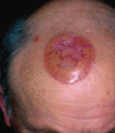
Figure 99.16
Necrobiotic xanthogranuloma. A plaque with yellowish hue involving the scalp.
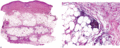
Figure 99.20
Histopathological features of a fully developed lesion of erythema nodosum. (a) Scanning power showing thickened septa of the subcutaneous tissue. (b)...
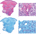
Figure 99.24
Histopathological features of erythema nodosum leprosum. (a) Scanning power showing dense nodular infiltrates in the dermis and a mostly lobular panni...
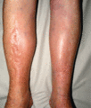
Figure 99.28
Late‐stage sclerosing panniculitis showing depressed hard areas with woody induration involving the lower legs.
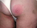
Figure 99.32
Erythematous nodule on gluteal region of a woman with equestrian cold panniculitis.
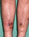
Figure 99.36
Pancreatic panniculitis in an alcoholic male. Nodular lesions, many of them ulcerated around the ankles.
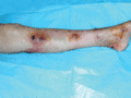
Figure 99.40
Sporotrichoid arrangement of subcutaneous nodules, several of them ulcerated and draining serous or oily discharge in an immunocompromised patient. Cu...
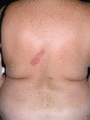
Figure 99.44
Neutrophilic lobular panniculitis showing erythematous plaques and nodules on the back of an adult woman.
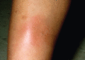
Figure 99.48
Traumatic panniculitis involving the shin.
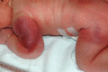
Figure 99.52
Subcutaneous fat necrosis of the newborn. Erythematous and violaceous plaques involving the shoulders, arms and buttocks.
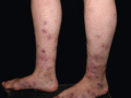
Figure 99.56
Gouty panniculitis. Ulcerated nodules on the lower legs of a patient with hyperuricaemia.
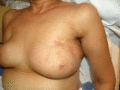
Figure 99.60
Sclerosing postirradiation panniculitis showing a depressed and indurate nodule on the previously irradiated area of the skin.
